Adriano de Oliveira Loures, Matheus de Abreu, Karina Lopes Devito, Eric Flavio Grisolia-Seifert, Katharina Jähn-Rickert, Gustavo Davi Rabelo
{"title":"下颌骨皮质骨的分形分析:根尖周X线片和显微计算机断层扫描与锥束计算机断层扫描两种处理方法获得的分形维数之间的相关性。","authors":"Adriano de Oliveira Loures, Matheus de Abreu, Karina Lopes Devito, Eric Flavio Grisolia-Seifert, Katharina Jähn-Rickert, Gustavo Davi Rabelo","doi":"10.1007/s00411-023-01045-0","DOIUrl":null,"url":null,"abstract":"<p><p>The objectives of the present study were to assess Fractal Dimension (FD) values in the mandible cortical bone obtained from digital periapical radiographs (DPR), high-resolution microtomography (µCT), and cone-beam computed tomography (CBCT), by two processing methods: binarization (FD.b) and grayscale-based method (FD.f) and, finally, to identify the correlation among these values with other micro-architectural parameters. For this, a prospective study was conducted on 18 healthy individuals (mean age 23 ± 2.4 years old) who underwent third molar extraction. Pre-operative CBCT scans were conducted, bone fragments were removed from the retro-molar region, and DPR and µCT were performed on those bone samples. FD.b and FD.f values were calculated using three parasagittal sections for CBCT, one image for DPR, and three sections for µCT. The 3D bone microarchitecture was analyzed in µCT (voxel size: 19 µm). As a result, FD.b mean values of 1.55 ± 0.02 and 1.80 ± 0.01 were obtained for CBCT and µCT, respectively. Furthermore, FD.f mean values of 1.22 ± 0.12 for DPR, 0.99 ± 0.04 for CBCT, and 1.30 ± 0.07 for µCT were obtained. Both FD.b and FD.f values showed a good agreement. FD.f was negatively correlated with the standard deviation of the mean gray value (p = 0.003) for DPR and intra-cortical bone surface (p = 0.02) for µCT. In conclusion, image processing with or without binarization revealed different values for FD, although showing agreement. The grayscale-based method retrieved FD values correlated with the gray levels and the cortical porous network, which means that FD can be a valuable index for mandibular cortical bone evaluation. FD is associated with mineralization and microarchitecture. Nevertheless, there was no correlation between FD values obtained from low- (DPR) and high-resolution (µCT) X-ray modalities with FD obtained from the in vivo CBCT.</p>","PeriodicalId":21002,"journal":{"name":"Radiation and Environmental Biophysics","volume":" ","pages":"511-518"},"PeriodicalIF":2.3000,"publicationDate":"2023-11-01","publicationTypes":"Journal Article","fieldsOfStudy":null,"isOpenAccess":false,"openAccessPdf":"","citationCount":"0","resultStr":"{\"title\":\"Fractal analysis of the mandible cortical bone: correlation among fractal dimension values obtained by two processing methods from periapical radiograph and micro-computed tomography with cone-beam computed tomography.\",\"authors\":\"Adriano de Oliveira Loures, Matheus de Abreu, Karina Lopes Devito, Eric Flavio Grisolia-Seifert, Katharina Jähn-Rickert, Gustavo Davi Rabelo\",\"doi\":\"10.1007/s00411-023-01045-0\",\"DOIUrl\":null,\"url\":null,\"abstract\":\"<p><p>The objectives of the present study were to assess Fractal Dimension (FD) values in the mandible cortical bone obtained from digital periapical radiographs (DPR), high-resolution microtomography (µCT), and cone-beam computed tomography (CBCT), by two processing methods: binarization (FD.b) and grayscale-based method (FD.f) and, finally, to identify the correlation among these values with other micro-architectural parameters. For this, a prospective study was conducted on 18 healthy individuals (mean age 23 ± 2.4 years old) who underwent third molar extraction. Pre-operative CBCT scans were conducted, bone fragments were removed from the retro-molar region, and DPR and µCT were performed on those bone samples. FD.b and FD.f values were calculated using three parasagittal sections for CBCT, one image for DPR, and three sections for µCT. The 3D bone microarchitecture was analyzed in µCT (voxel size: 19 µm). As a result, FD.b mean values of 1.55 ± 0.02 and 1.80 ± 0.01 were obtained for CBCT and µCT, respectively. Furthermore, FD.f mean values of 1.22 ± 0.12 for DPR, 0.99 ± 0.04 for CBCT, and 1.30 ± 0.07 for µCT were obtained. Both FD.b and FD.f values showed a good agreement. FD.f was negatively correlated with the standard deviation of the mean gray value (p = 0.003) for DPR and intra-cortical bone surface (p = 0.02) for µCT. In conclusion, image processing with or without binarization revealed different values for FD, although showing agreement. The grayscale-based method retrieved FD values correlated with the gray levels and the cortical porous network, which means that FD can be a valuable index for mandibular cortical bone evaluation. FD is associated with mineralization and microarchitecture. Nevertheless, there was no correlation between FD values obtained from low- (DPR) and high-resolution (µCT) X-ray modalities with FD obtained from the in vivo CBCT.</p>\",\"PeriodicalId\":21002,\"journal\":{\"name\":\"Radiation and Environmental Biophysics\",\"volume\":\" \",\"pages\":\"511-518\"},\"PeriodicalIF\":2.3000,\"publicationDate\":\"2023-11-01\",\"publicationTypes\":\"Journal Article\",\"fieldsOfStudy\":null,\"isOpenAccess\":false,\"openAccessPdf\":\"\",\"citationCount\":\"0\",\"resultStr\":null,\"platform\":\"Semanticscholar\",\"paperid\":null,\"PeriodicalName\":\"Radiation and Environmental Biophysics\",\"FirstCategoryId\":\"93\",\"ListUrlMain\":\"https://doi.org/10.1007/s00411-023-01045-0\",\"RegionNum\":4,\"RegionCategory\":\"环境科学与生态学\",\"ArticlePicture\":[],\"TitleCN\":null,\"AbstractTextCN\":null,\"PMCID\":null,\"EPubDate\":\"2023/10/4 0:00:00\",\"PubModel\":\"Epub\",\"JCR\":\"Q3\",\"JCRName\":\"BIOLOGY\",\"Score\":null,\"Total\":0}","platform":"Semanticscholar","paperid":null,"PeriodicalName":"Radiation and Environmental Biophysics","FirstCategoryId":"93","ListUrlMain":"https://doi.org/10.1007/s00411-023-01045-0","RegionNum":4,"RegionCategory":"环境科学与生态学","ArticlePicture":[],"TitleCN":null,"AbstractTextCN":null,"PMCID":null,"EPubDate":"2023/10/4 0:00:00","PubModel":"Epub","JCR":"Q3","JCRName":"BIOLOGY","Score":null,"Total":0}
Fractal analysis of the mandible cortical bone: correlation among fractal dimension values obtained by two processing methods from periapical radiograph and micro-computed tomography with cone-beam computed tomography.
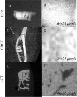
The objectives of the present study were to assess Fractal Dimension (FD) values in the mandible cortical bone obtained from digital periapical radiographs (DPR), high-resolution microtomography (µCT), and cone-beam computed tomography (CBCT), by two processing methods: binarization (FD.b) and grayscale-based method (FD.f) and, finally, to identify the correlation among these values with other micro-architectural parameters. For this, a prospective study was conducted on 18 healthy individuals (mean age 23 ± 2.4 years old) who underwent third molar extraction. Pre-operative CBCT scans were conducted, bone fragments were removed from the retro-molar region, and DPR and µCT were performed on those bone samples. FD.b and FD.f values were calculated using three parasagittal sections for CBCT, one image for DPR, and three sections for µCT. The 3D bone microarchitecture was analyzed in µCT (voxel size: 19 µm). As a result, FD.b mean values of 1.55 ± 0.02 and 1.80 ± 0.01 were obtained for CBCT and µCT, respectively. Furthermore, FD.f mean values of 1.22 ± 0.12 for DPR, 0.99 ± 0.04 for CBCT, and 1.30 ± 0.07 for µCT were obtained. Both FD.b and FD.f values showed a good agreement. FD.f was negatively correlated with the standard deviation of the mean gray value (p = 0.003) for DPR and intra-cortical bone surface (p = 0.02) for µCT. In conclusion, image processing with or without binarization revealed different values for FD, although showing agreement. The grayscale-based method retrieved FD values correlated with the gray levels and the cortical porous network, which means that FD can be a valuable index for mandibular cortical bone evaluation. FD is associated with mineralization and microarchitecture. Nevertheless, there was no correlation between FD values obtained from low- (DPR) and high-resolution (µCT) X-ray modalities with FD obtained from the in vivo CBCT.
期刊介绍:
This journal is devoted to fundamental and applied issues in radiation research and biophysics. The topics may include:
Biophysics of ionizing radiation: radiation physics and chemistry, radiation dosimetry, radiobiology, radioecology, biophysical foundations of medical applications of radiation, and radiation protection.
Biological effects of radiation: experimental or theoretical work on molecular or cellular effects; relevance of biological effects for risk assessment; biological effects of medical applications of radiation; relevance of radiation for biosphere and in space; modelling of ecosystems; modelling of transport processes of substances in biotic systems.
Risk assessment: epidemiological studies of cancer and non-cancer effects; quantification of risk including exposures to radiation and confounding factors
Contributions to these topics may include theoretical-mathematical and experimental material, as well as description of new techniques relevant for the study of these issues. They can range from complex radiobiological phenomena to issues in health physics and environmental protection.
 求助内容:
求助内容: 应助结果提醒方式:


