Magdalena Walewska, Anna Małek, Bartosz Taciak, Anna Wojtalewicz, Sylwia Wilk, Agata Wojtkowska, Katarzyna Zabielska-Koczywąs, Roman Lechowski
{"title":"聚乙二醇脂质体阿霉素作为犬转移性骨肉瘤的潜在治疗剂——体外和卵外研究。","authors":"Magdalena Walewska, Anna Małek, Bartosz Taciak, Anna Wojtalewicz, Sylwia Wilk, Agata Wojtkowska, Katarzyna Zabielska-Koczywąs, Roman Lechowski","doi":"10.2478/jvetres-2023-0026","DOIUrl":null,"url":null,"abstract":"<p><strong>Introduction: </strong>Appendicular osteosarcoma (OSA) is a highly aggressive and metastatic primary bone tumour in dogs. Standard therapy is amputation and adjuvant chemotherapy (<i>e.g</i>. with doxorubicin). Liposomal drug delivery may augment therapeutic efficacy and reduce negative side effects. Polyethylene glycol (PEG)-liposomal doxorubicin treats human metastatic cancers effectively. The study aimed was to evaluate PEG-liposomal doxorubicin's inhibitory effect on canine metastatic proliferation and migration <i>in vitro</i>. It also aimed to appraise the drug's extravasation inhibition <i>in vivo</i> using the human medicine-proven chick embryo chorioallantoic membrane <i>ex ovo</i> model.</p><p><strong>Material and methods: </strong>The canine D-17 OSA cell line was cultured and inoculated with decreasing concentrations of PEG-liposomal doxorubicin and conventional doxorubicin in a 3-(4, 5-dimethylthiazol-2-yl)-2, 5-diphenyltetrazolium bromide (MTT) test of cell viability, proliferation and cytotoxicity. Flow cytometry with Annexin V and Draq 7 staining confirmed the MTT test results, indicating dead, early and late apoptotic, and live cells. The inhibitory effect of the two preparations on cancer cell migration was investigated with a wound-healing assay. Culture plates seeded with cells were prepared. The cell monolayer was scratched and images of cells migrating to the scratch were captured at 0 h, 12 h and 24 h. Also, embryos were removed from three-day-incubated fertilised chicken eggs. On the 12<sup>th</sup> day, labelled D-17 cells were injected into each embryo. Embryos in one group received 100 μL of phosphate-buffered saline as controls, those in another group 30 μg/mL of PEG-liposomal doxorubicin, and those in the last group 6 μg/mL of conventional doxorubicin. The effectiveness of the intravascular administration of the D-17 cells was confirmed under a microscope.</p><p><strong>Results: </strong>PEG-liposomal doxorubicin inhibited the migration of canine OSA cells more effectively than conventional doxorubicin (P ≤ 0.05). The <i>ex ovo</i> model showed that both drugs had similar impacts on canine metastatic OSA.</p><p><strong>Conclusion: </strong>The liposomal form of the drug may be considered a potentially effective compound in canine metastatic OSA; nevertheless, further <i>in vivo</i> studies are essential to confirm this hypothesis.</p>","PeriodicalId":17617,"journal":{"name":"Journal of Veterinary Research","volume":"67 2","pages":"297-305"},"PeriodicalIF":1.3000,"publicationDate":"2023-06-16","publicationTypes":"Journal Article","fieldsOfStudy":null,"isOpenAccess":false,"openAccessPdf":"https://ftp.ncbi.nlm.nih.gov/pub/pmc/oa_pdf/42/ef/jvetres-67-2-jvetres-2023-0026.PMC10541662.pdf","citationCount":"0","resultStr":"{\"title\":\"PEG-liposomal doxorubicin as a potential agent for canine metastatic osteosarcoma - <i>in vitro</i> and <i>ex ovo</i> studies.\",\"authors\":\"Magdalena Walewska, Anna Małek, Bartosz Taciak, Anna Wojtalewicz, Sylwia Wilk, Agata Wojtkowska, Katarzyna Zabielska-Koczywąs, Roman Lechowski\",\"doi\":\"10.2478/jvetres-2023-0026\",\"DOIUrl\":null,\"url\":null,\"abstract\":\"<p><strong>Introduction: </strong>Appendicular osteosarcoma (OSA) is a highly aggressive and metastatic primary bone tumour in dogs. Standard therapy is amputation and adjuvant chemotherapy (<i>e.g</i>. with doxorubicin). Liposomal drug delivery may augment therapeutic efficacy and reduce negative side effects. Polyethylene glycol (PEG)-liposomal doxorubicin treats human metastatic cancers effectively. The study aimed was to evaluate PEG-liposomal doxorubicin's inhibitory effect on canine metastatic proliferation and migration <i>in vitro</i>. It also aimed to appraise the drug's extravasation inhibition <i>in vivo</i> using the human medicine-proven chick embryo chorioallantoic membrane <i>ex ovo</i> model.</p><p><strong>Material and methods: </strong>The canine D-17 OSA cell line was cultured and inoculated with decreasing concentrations of PEG-liposomal doxorubicin and conventional doxorubicin in a 3-(4, 5-dimethylthiazol-2-yl)-2, 5-diphenyltetrazolium bromide (MTT) test of cell viability, proliferation and cytotoxicity. Flow cytometry with Annexin V and Draq 7 staining confirmed the MTT test results, indicating dead, early and late apoptotic, and live cells. The inhibitory effect of the two preparations on cancer cell migration was investigated with a wound-healing assay. Culture plates seeded with cells were prepared. The cell monolayer was scratched and images of cells migrating to the scratch were captured at 0 h, 12 h and 24 h. Also, embryos were removed from three-day-incubated fertilised chicken eggs. On the 12<sup>th</sup> day, labelled D-17 cells were injected into each embryo. Embryos in one group received 100 μL of phosphate-buffered saline as controls, those in another group 30 μg/mL of PEG-liposomal doxorubicin, and those in the last group 6 μg/mL of conventional doxorubicin. The effectiveness of the intravascular administration of the D-17 cells was confirmed under a microscope.</p><p><strong>Results: </strong>PEG-liposomal doxorubicin inhibited the migration of canine OSA cells more effectively than conventional doxorubicin (P ≤ 0.05). The <i>ex ovo</i> model showed that both drugs had similar impacts on canine metastatic OSA.</p><p><strong>Conclusion: </strong>The liposomal form of the drug may be considered a potentially effective compound in canine metastatic OSA; nevertheless, further <i>in vivo</i> studies are essential to confirm this hypothesis.</p>\",\"PeriodicalId\":17617,\"journal\":{\"name\":\"Journal of Veterinary Research\",\"volume\":\"67 2\",\"pages\":\"297-305\"},\"PeriodicalIF\":1.3000,\"publicationDate\":\"2023-06-16\",\"publicationTypes\":\"Journal Article\",\"fieldsOfStudy\":null,\"isOpenAccess\":false,\"openAccessPdf\":\"https://ftp.ncbi.nlm.nih.gov/pub/pmc/oa_pdf/42/ef/jvetres-67-2-jvetres-2023-0026.PMC10541662.pdf\",\"citationCount\":\"0\",\"resultStr\":null,\"platform\":\"Semanticscholar\",\"paperid\":null,\"PeriodicalName\":\"Journal of Veterinary Research\",\"FirstCategoryId\":\"97\",\"ListUrlMain\":\"https://doi.org/10.2478/jvetres-2023-0026\",\"RegionNum\":3,\"RegionCategory\":\"农林科学\",\"ArticlePicture\":[],\"TitleCN\":null,\"AbstractTextCN\":null,\"PMCID\":null,\"EPubDate\":\"2023/6/1 0:00:00\",\"PubModel\":\"eCollection\",\"JCR\":\"Q2\",\"JCRName\":\"VETERINARY SCIENCES\",\"Score\":null,\"Total\":0}","platform":"Semanticscholar","paperid":null,"PeriodicalName":"Journal of Veterinary Research","FirstCategoryId":"97","ListUrlMain":"https://doi.org/10.2478/jvetres-2023-0026","RegionNum":3,"RegionCategory":"农林科学","ArticlePicture":[],"TitleCN":null,"AbstractTextCN":null,"PMCID":null,"EPubDate":"2023/6/1 0:00:00","PubModel":"eCollection","JCR":"Q2","JCRName":"VETERINARY SCIENCES","Score":null,"Total":0}
PEG-liposomal doxorubicin as a potential agent for canine metastatic osteosarcoma - in vitro and ex ovo studies.
Introduction: Appendicular osteosarcoma (OSA) is a highly aggressive and metastatic primary bone tumour in dogs. Standard therapy is amputation and adjuvant chemotherapy (e.g. with doxorubicin). Liposomal drug delivery may augment therapeutic efficacy and reduce negative side effects. Polyethylene glycol (PEG)-liposomal doxorubicin treats human metastatic cancers effectively. The study aimed was to evaluate PEG-liposomal doxorubicin's inhibitory effect on canine metastatic proliferation and migration in vitro. It also aimed to appraise the drug's extravasation inhibition in vivo using the human medicine-proven chick embryo chorioallantoic membrane ex ovo model.
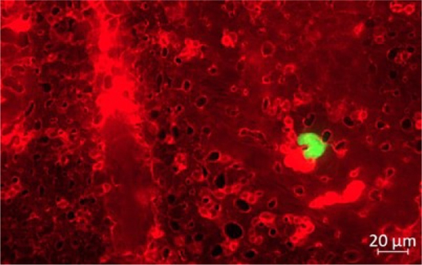
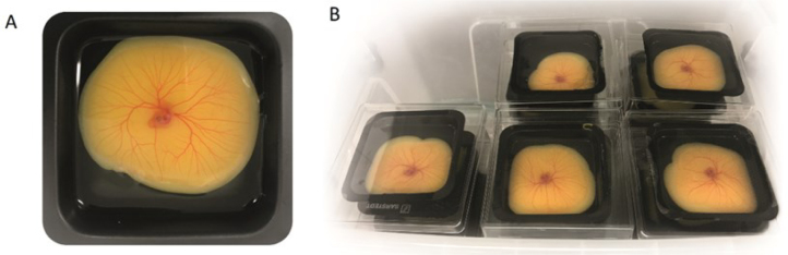
Material and methods: The canine D-17 OSA cell line was cultured and inoculated with decreasing concentrations of PEG-liposomal doxorubicin and conventional doxorubicin in a 3-(4, 5-dimethylthiazol-2-yl)-2, 5-diphenyltetrazolium bromide (MTT) test of cell viability, proliferation and cytotoxicity. Flow cytometry with Annexin V and Draq 7 staining confirmed the MTT test results, indicating dead, early and late apoptotic, and live cells. The inhibitory effect of the two preparations on cancer cell migration was investigated with a wound-healing assay. Culture plates seeded with cells were prepared. The cell monolayer was scratched and images of cells migrating to the scratch were captured at 0 h, 12 h and 24 h. Also, embryos were removed from three-day-incubated fertilised chicken eggs. On the 12th day, labelled D-17 cells were injected into each embryo. Embryos in one group received 100 μL of phosphate-buffered saline as controls, those in another group 30 μg/mL of PEG-liposomal doxorubicin, and those in the last group 6 μg/mL of conventional doxorubicin. The effectiveness of the intravascular administration of the D-17 cells was confirmed under a microscope.
Results: PEG-liposomal doxorubicin inhibited the migration of canine OSA cells more effectively than conventional doxorubicin (P ≤ 0.05). The ex ovo model showed that both drugs had similar impacts on canine metastatic OSA.
Conclusion: The liposomal form of the drug may be considered a potentially effective compound in canine metastatic OSA; nevertheless, further in vivo studies are essential to confirm this hypothesis.
期刊介绍:
Journal of Veterinary Research (formerly Bulletin of the Veterinary Institute in Pulawy) is a quarterly that publishes original papers, review articles and short communications on bacteriology, virology, parasitology, immunology, molecular biology, pathology, toxicology, pharmacology, and biochemistry. The main emphasis is, however, on infectious diseases of animals, food safety and public health, and clinical sciences.

 求助内容:
求助内容: 应助结果提醒方式:


