{"title":"一名西班牙裔患者的非典型Mal de Meleda。","authors":"Mónica Guevara, Michelle Mafla, Camila Miño","doi":"10.1155/2023/6640311","DOIUrl":null,"url":null,"abstract":"<p><p>Mal de Meleda (MDM) is a rare autosomal palmoplantar keratoderma (PPK) skin disorder (estimated incidence of 1 per 100,000 people) commonly associated with consanguinity and early childhood onset. MDM is characterized by bilateral diffusion of PPK plaques with delimited yellowish lesions that transgredien to the dorsum of the hands and feet. Additional features include nail dystrophy, lichenoid lesions, hyperhidrotic maceration, involvement of the knees and elbows, malodor, fungal superinfections, and digital constrictions. A male patient aged 42 years presented with asymptomatic, chronic, and diffused PPK lesions that progressed to the dorsal surface of the hands and feet, along with knees and elbows involvement. On clinical examination, asymmetrical lesions were observed on the hands, the left palm with yellowish waxy hyperkeratotic plaques, and the right palm with erythematous scaling and hyperkeratotic interphalangeal rings. The soles of the feet presented with yellow waxy hyperkeratotic plaques. In addition, nail dystrophy and loss of dermatoglyphics were observed. Initially, symptomatic topical treatment was established. However, owing to the lack of clinical response, a biopsy was performed, which revealed thickened corneal layer, acanthosis, spongiosis, and perivascular lymphohistiocytic infiltrate. MDM diagnosis was confirmed based on a personal history of consanguinity, clinical presentation with absence of systemic symptoms, and transgredien pattern of the lesions. Systemic treatment with low doses of isotretinoin (10 mg orally everyday) was initiated, and two months later, slight clinical improvement has been observed until date. The present case report describes MDM in a Hispanic patient, who presented with asymmetric PPK lesions on the hands and received isotretinoin treatment.</p>","PeriodicalId":9630,"journal":{"name":"Case Reports in Dermatological Medicine","volume":"2023 ","pages":"6640311"},"PeriodicalIF":0.0000,"publicationDate":"2023-09-14","publicationTypes":"Journal Article","fieldsOfStudy":null,"isOpenAccess":false,"openAccessPdf":"https://www.ncbi.nlm.nih.gov/pmc/articles/PMC10513804/pdf/","citationCount":"0","resultStr":"{\"title\":\"Atypical Mal de Meleda in a Hispanic Patient.\",\"authors\":\"Mónica Guevara, Michelle Mafla, Camila Miño\",\"doi\":\"10.1155/2023/6640311\",\"DOIUrl\":null,\"url\":null,\"abstract\":\"<p><p>Mal de Meleda (MDM) is a rare autosomal palmoplantar keratoderma (PPK) skin disorder (estimated incidence of 1 per 100,000 people) commonly associated with consanguinity and early childhood onset. MDM is characterized by bilateral diffusion of PPK plaques with delimited yellowish lesions that transgredien to the dorsum of the hands and feet. Additional features include nail dystrophy, lichenoid lesions, hyperhidrotic maceration, involvement of the knees and elbows, malodor, fungal superinfections, and digital constrictions. A male patient aged 42 years presented with asymptomatic, chronic, and diffused PPK lesions that progressed to the dorsal surface of the hands and feet, along with knees and elbows involvement. On clinical examination, asymmetrical lesions were observed on the hands, the left palm with yellowish waxy hyperkeratotic plaques, and the right palm with erythematous scaling and hyperkeratotic interphalangeal rings. The soles of the feet presented with yellow waxy hyperkeratotic plaques. In addition, nail dystrophy and loss of dermatoglyphics were observed. Initially, symptomatic topical treatment was established. However, owing to the lack of clinical response, a biopsy was performed, which revealed thickened corneal layer, acanthosis, spongiosis, and perivascular lymphohistiocytic infiltrate. MDM diagnosis was confirmed based on a personal history of consanguinity, clinical presentation with absence of systemic symptoms, and transgredien pattern of the lesions. Systemic treatment with low doses of isotretinoin (10 mg orally everyday) was initiated, and two months later, slight clinical improvement has been observed until date. The present case report describes MDM in a Hispanic patient, who presented with asymmetric PPK lesions on the hands and received isotretinoin treatment.</p>\",\"PeriodicalId\":9630,\"journal\":{\"name\":\"Case Reports in Dermatological Medicine\",\"volume\":\"2023 \",\"pages\":\"6640311\"},\"PeriodicalIF\":0.0000,\"publicationDate\":\"2023-09-14\",\"publicationTypes\":\"Journal Article\",\"fieldsOfStudy\":null,\"isOpenAccess\":false,\"openAccessPdf\":\"https://www.ncbi.nlm.nih.gov/pmc/articles/PMC10513804/pdf/\",\"citationCount\":\"0\",\"resultStr\":null,\"platform\":\"Semanticscholar\",\"paperid\":null,\"PeriodicalName\":\"Case Reports in Dermatological Medicine\",\"FirstCategoryId\":\"1085\",\"ListUrlMain\":\"https://doi.org/10.1155/2023/6640311\",\"RegionNum\":0,\"RegionCategory\":null,\"ArticlePicture\":[],\"TitleCN\":null,\"AbstractTextCN\":null,\"PMCID\":null,\"EPubDate\":\"2023/1/1 0:00:00\",\"PubModel\":\"eCollection\",\"JCR\":\"Q3\",\"JCRName\":\"Medicine\",\"Score\":null,\"Total\":0}","platform":"Semanticscholar","paperid":null,"PeriodicalName":"Case Reports in Dermatological Medicine","FirstCategoryId":"1085","ListUrlMain":"https://doi.org/10.1155/2023/6640311","RegionNum":0,"RegionCategory":null,"ArticlePicture":[],"TitleCN":null,"AbstractTextCN":null,"PMCID":null,"EPubDate":"2023/1/1 0:00:00","PubModel":"eCollection","JCR":"Q3","JCRName":"Medicine","Score":null,"Total":0}
引用次数: 0
摘要
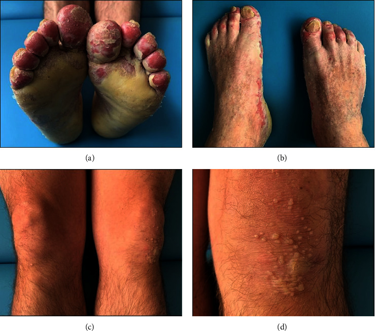
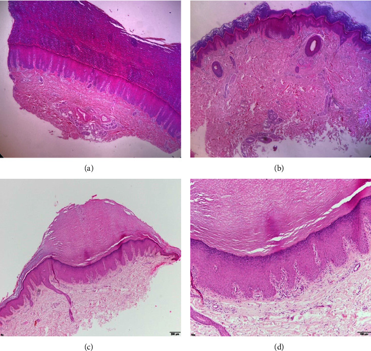
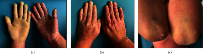
Mal de Meleda(MDM)是一种罕见的常染色体掌跖角化病(PPK)皮肤病(估计发病率为十万分之一),通常与血缘关系和儿童早期发病有关。MDM的特征是PPK斑块的双侧扩散,有界限的黄色病变,这些病变转移到手和脚的背侧。其他特征包括指甲营养不良、地衣样病变、多汗浸渍、膝盖和肘部受累、恶臭、真菌重叠感染和指关节收缩。一名42岁的男性患者 年表现为无症状、慢性和弥漫性PPK病变,进展至手和脚的背表面,同时伴有膝盖和肘部受累。临床检查发现手部不对称病变,左手掌有黄蜡状角化过度斑块,右手掌有红斑性鳞屑和角化过度的指间环。脚底出现黄色蜡状角化过度斑块。此外,还观察到指甲营养不良和皮纹缺失。最初,建立了症状性局部治疗。然而,由于缺乏临床反应,进行了活检,发现角膜层增厚、棘皮病、海绵状血管病和血管周围淋巴组织细胞浸润。MDM的诊断是根据个人血亲史、无全身症状的临床表现和病变的变性模式来确认的。低剂量异维甲酸的全身治疗(10 每日口服mg),两个月后,迄今为止,观察到轻微的临床改善。本病例报告描述了一名西班牙裔患者的MDM,该患者手部出现不对称PPK病变,并接受了异维甲酸治疗。
Mal de Meleda (MDM) is a rare autosomal palmoplantar keratoderma (PPK) skin disorder (estimated incidence of 1 per 100,000 people) commonly associated with consanguinity and early childhood onset. MDM is characterized by bilateral diffusion of PPK plaques with delimited yellowish lesions that transgredien to the dorsum of the hands and feet. Additional features include nail dystrophy, lichenoid lesions, hyperhidrotic maceration, involvement of the knees and elbows, malodor, fungal superinfections, and digital constrictions. A male patient aged 42 years presented with asymptomatic, chronic, and diffused PPK lesions that progressed to the dorsal surface of the hands and feet, along with knees and elbows involvement. On clinical examination, asymmetrical lesions were observed on the hands, the left palm with yellowish waxy hyperkeratotic plaques, and the right palm with erythematous scaling and hyperkeratotic interphalangeal rings. The soles of the feet presented with yellow waxy hyperkeratotic plaques. In addition, nail dystrophy and loss of dermatoglyphics were observed. Initially, symptomatic topical treatment was established. However, owing to the lack of clinical response, a biopsy was performed, which revealed thickened corneal layer, acanthosis, spongiosis, and perivascular lymphohistiocytic infiltrate. MDM diagnosis was confirmed based on a personal history of consanguinity, clinical presentation with absence of systemic symptoms, and transgredien pattern of the lesions. Systemic treatment with low doses of isotretinoin (10 mg orally everyday) was initiated, and two months later, slight clinical improvement has been observed until date. The present case report describes MDM in a Hispanic patient, who presented with asymmetric PPK lesions on the hands and received isotretinoin treatment.

 求助内容:
求助内容: 应助结果提醒方式:


