E M Trubushkina, E M Boyko, D V Stomatov, I V Rzhepakovsky, S I Piskov, D S-A Yeldashev, A A Kutsenko, A A Dolgalev
{"title":"上颌窦前壁骨重建方法的比较(实验研究)。","authors":"E M Trubushkina, E M Boyko, D V Stomatov, I V Rzhepakovsky, S I Piskov, D S-A Yeldashev, A A Kutsenko, A A Dolgalev","doi":"10.17691/stm2022.14.1.05","DOIUrl":null,"url":null,"abstract":"<p><p><b>The aim of the study</b> was to compare various methods used for the bone reconstruction in the anterior wall of the maxillary sinus during sinus lift surgery; in addition, we aimed to study the effect of maxillary sinus membrane perforation on the healing process.</p><p><strong>Materials and methods: </strong>The experiments were carried out using the North Caucasian sheep. Maxillary sinus lift surgery was performed on the animals under general anesthesia. The skin and muscle fascia were dissected layer-by-layer providing the optimal conditions for bone preparation; then, three bone windows were made on each side of the head. Two windows were sawn out with a spherical bur, the third window - with a hollow bur and part of the anterior wall was taken out. On one side, the mucous membrane of the maxillary sinus was pulled and perforated; on the other side, the sinus lift was performed with no membrane perforation. On each side, one window was left uncovered, the second was closed with a collagen membrane, and the third was closed with a bone cover. After 30 and 60 days, the sheep were taken out of the experiment in groups of three; samples were collected from the operated areas and examined using computed microtomography and histology.</p><p><strong>Results: </strong>According to the histological study, the bone repair process developed normally regardless of the surgery technique. The process started with the appearance of granulation tissue and connective tissue cords; in the final stages, cellular differentiation, pronounced osteoblastic activity, and inter-beam formation were seen.The most active regeneration was observed in the areas where the bone defects were closed with a collagen membrane, and especially in the windows made with no perforation of the maxillary sinus membrane. The microtomographic and histological tests proved that perforation of the mucous membrane during the sinus lift operation impaired bone tissue regeneration.</p><p><strong>Conclusion: </strong>The obtained results suggest that the most promising way to close a bone defect in the anterior wall of the maxillary sinus is the use of a collagen membrane; therefore, we recommend choosing this approach for sinus lift surgery.</p>","PeriodicalId":51886,"journal":{"name":"Sovremennye Tehnologii v Medicine","volume":"14 1","pages":"44-53"},"PeriodicalIF":0.9000,"publicationDate":"2022-01-01","publicationTypes":"Journal Article","fieldsOfStudy":null,"isOpenAccess":false,"openAccessPdf":"https://www.ncbi.nlm.nih.gov/pmc/articles/PMC9376757/pdf/","citationCount":"0","resultStr":"{\"title\":\"The Comparison of Methods for Bone Reconstruction in the Anterior Wall of the Maxillary Sinus (an Experimental Study).\",\"authors\":\"E M Trubushkina, E M Boyko, D V Stomatov, I V Rzhepakovsky, S I Piskov, D S-A Yeldashev, A A Kutsenko, A A Dolgalev\",\"doi\":\"10.17691/stm2022.14.1.05\",\"DOIUrl\":null,\"url\":null,\"abstract\":\"<p><p><b>The aim of the study</b> was to compare various methods used for the bone reconstruction in the anterior wall of the maxillary sinus during sinus lift surgery; in addition, we aimed to study the effect of maxillary sinus membrane perforation on the healing process.</p><p><strong>Materials and methods: </strong>The experiments were carried out using the North Caucasian sheep. Maxillary sinus lift surgery was performed on the animals under general anesthesia. The skin and muscle fascia were dissected layer-by-layer providing the optimal conditions for bone preparation; then, three bone windows were made on each side of the head. Two windows were sawn out with a spherical bur, the third window - with a hollow bur and part of the anterior wall was taken out. On one side, the mucous membrane of the maxillary sinus was pulled and perforated; on the other side, the sinus lift was performed with no membrane perforation. On each side, one window was left uncovered, the second was closed with a collagen membrane, and the third was closed with a bone cover. After 30 and 60 days, the sheep were taken out of the experiment in groups of three; samples were collected from the operated areas and examined using computed microtomography and histology.</p><p><strong>Results: </strong>According to the histological study, the bone repair process developed normally regardless of the surgery technique. The process started with the appearance of granulation tissue and connective tissue cords; in the final stages, cellular differentiation, pronounced osteoblastic activity, and inter-beam formation were seen.The most active regeneration was observed in the areas where the bone defects were closed with a collagen membrane, and especially in the windows made with no perforation of the maxillary sinus membrane. The microtomographic and histological tests proved that perforation of the mucous membrane during the sinus lift operation impaired bone tissue regeneration.</p><p><strong>Conclusion: </strong>The obtained results suggest that the most promising way to close a bone defect in the anterior wall of the maxillary sinus is the use of a collagen membrane; therefore, we recommend choosing this approach for sinus lift surgery.</p>\",\"PeriodicalId\":51886,\"journal\":{\"name\":\"Sovremennye Tehnologii v Medicine\",\"volume\":\"14 1\",\"pages\":\"44-53\"},\"PeriodicalIF\":0.9000,\"publicationDate\":\"2022-01-01\",\"publicationTypes\":\"Journal Article\",\"fieldsOfStudy\":null,\"isOpenAccess\":false,\"openAccessPdf\":\"https://www.ncbi.nlm.nih.gov/pmc/articles/PMC9376757/pdf/\",\"citationCount\":\"0\",\"resultStr\":null,\"platform\":\"Semanticscholar\",\"paperid\":null,\"PeriodicalName\":\"Sovremennye Tehnologii v Medicine\",\"FirstCategoryId\":\"1085\",\"ListUrlMain\":\"https://doi.org/10.17691/stm2022.14.1.05\",\"RegionNum\":0,\"RegionCategory\":null,\"ArticlePicture\":[],\"TitleCN\":null,\"AbstractTextCN\":null,\"PMCID\":null,\"EPubDate\":\"2022/1/28 0:00:00\",\"PubModel\":\"Epub\",\"JCR\":\"Q4\",\"JCRName\":\"MEDICINE, RESEARCH & EXPERIMENTAL\",\"Score\":null,\"Total\":0}","platform":"Semanticscholar","paperid":null,"PeriodicalName":"Sovremennye Tehnologii v Medicine","FirstCategoryId":"1085","ListUrlMain":"https://doi.org/10.17691/stm2022.14.1.05","RegionNum":0,"RegionCategory":null,"ArticlePicture":[],"TitleCN":null,"AbstractTextCN":null,"PMCID":null,"EPubDate":"2022/1/28 0:00:00","PubModel":"Epub","JCR":"Q4","JCRName":"MEDICINE, RESEARCH & EXPERIMENTAL","Score":null,"Total":0}
The Comparison of Methods for Bone Reconstruction in the Anterior Wall of the Maxillary Sinus (an Experimental Study).
The aim of the study was to compare various methods used for the bone reconstruction in the anterior wall of the maxillary sinus during sinus lift surgery; in addition, we aimed to study the effect of maxillary sinus membrane perforation on the healing process.
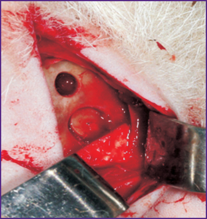
Materials and methods: The experiments were carried out using the North Caucasian sheep. Maxillary sinus lift surgery was performed on the animals under general anesthesia. The skin and muscle fascia were dissected layer-by-layer providing the optimal conditions for bone preparation; then, three bone windows were made on each side of the head. Two windows were sawn out with a spherical bur, the third window - with a hollow bur and part of the anterior wall was taken out. On one side, the mucous membrane of the maxillary sinus was pulled and perforated; on the other side, the sinus lift was performed with no membrane perforation. On each side, one window was left uncovered, the second was closed with a collagen membrane, and the third was closed with a bone cover. After 30 and 60 days, the sheep were taken out of the experiment in groups of three; samples were collected from the operated areas and examined using computed microtomography and histology.
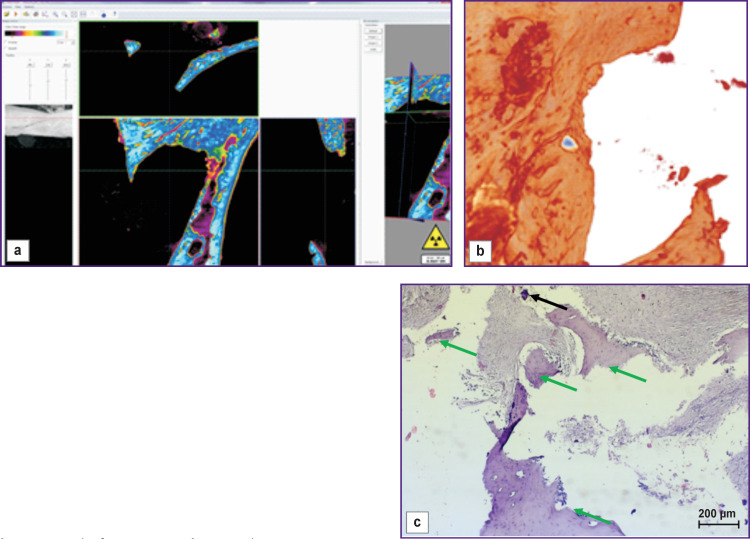
Results: According to the histological study, the bone repair process developed normally regardless of the surgery technique. The process started with the appearance of granulation tissue and connective tissue cords; in the final stages, cellular differentiation, pronounced osteoblastic activity, and inter-beam formation were seen.The most active regeneration was observed in the areas where the bone defects were closed with a collagen membrane, and especially in the windows made with no perforation of the maxillary sinus membrane. The microtomographic and histological tests proved that perforation of the mucous membrane during the sinus lift operation impaired bone tissue regeneration.
Conclusion: The obtained results suggest that the most promising way to close a bone defect in the anterior wall of the maxillary sinus is the use of a collagen membrane; therefore, we recommend choosing this approach for sinus lift surgery.


 求助内容:
求助内容: 应助结果提醒方式:


