Theofanis George Korovesis, Paraskevi Koutrolou-Sotiropoulou, Demosthenes George Katritsis
{"title":"致心律失常二尖瓣脱垂。","authors":"Theofanis George Korovesis, Paraskevi Koutrolou-Sotiropoulou, Demosthenes George Katritsis","doi":"10.15420/aer.2021.28","DOIUrl":null,"url":null,"abstract":"<p><p>Mitral valve prolapse (MVP) is a common condition present in 1-3% of the population. There has been evidence that a subset of MVP patients is at higher risk of sudden cardiac death. The arrhythmogenic mechanism is related to fibrotic changes in the papillary muscles caused by the prolapsing valve. ECG features include ST-segment depression, T wave inversion or biphasic T waves in inferior leads, and premature ventricular contractions arising from the papillary muscles and the fascicular system. Echocardiography can identify MVP and mitral annular disjunction, a feature that has significant negative prognostic value in MVP. Cardiac MRI is indicated for identifying fibrosis. Patients with high-risk features should be referred for further evaluation. Catheter ablation and mitral valve repair might reduce the risk of malignant arrhythmia. MVP patients with high-risk features and clinically documented ventricular arrhythmia may also be considered for an ICD.</p>","PeriodicalId":8412,"journal":{"name":"Arrhythmia & Electrophysiology Review","volume":" ","pages":"e16"},"PeriodicalIF":3.3000,"publicationDate":"2022-04-01","publicationTypes":"Journal Article","fieldsOfStudy":null,"isOpenAccess":false,"openAccessPdf":"https://ftp.ncbi.nlm.nih.gov/pub/pmc/oa_pdf/e7/e3/aer-11-e16.PMC9376835.pdf","citationCount":"2","resultStr":"{\"title\":\"Arrhythmogenic Mitral Valve Prolapse.\",\"authors\":\"Theofanis George Korovesis, Paraskevi Koutrolou-Sotiropoulou, Demosthenes George Katritsis\",\"doi\":\"10.15420/aer.2021.28\",\"DOIUrl\":null,\"url\":null,\"abstract\":\"<p><p>Mitral valve prolapse (MVP) is a common condition present in 1-3% of the population. There has been evidence that a subset of MVP patients is at higher risk of sudden cardiac death. The arrhythmogenic mechanism is related to fibrotic changes in the papillary muscles caused by the prolapsing valve. ECG features include ST-segment depression, T wave inversion or biphasic T waves in inferior leads, and premature ventricular contractions arising from the papillary muscles and the fascicular system. Echocardiography can identify MVP and mitral annular disjunction, a feature that has significant negative prognostic value in MVP. Cardiac MRI is indicated for identifying fibrosis. Patients with high-risk features should be referred for further evaluation. Catheter ablation and mitral valve repair might reduce the risk of malignant arrhythmia. MVP patients with high-risk features and clinically documented ventricular arrhythmia may also be considered for an ICD.</p>\",\"PeriodicalId\":8412,\"journal\":{\"name\":\"Arrhythmia & Electrophysiology Review\",\"volume\":\" \",\"pages\":\"e16\"},\"PeriodicalIF\":3.3000,\"publicationDate\":\"2022-04-01\",\"publicationTypes\":\"Journal Article\",\"fieldsOfStudy\":null,\"isOpenAccess\":false,\"openAccessPdf\":\"https://ftp.ncbi.nlm.nih.gov/pub/pmc/oa_pdf/e7/e3/aer-11-e16.PMC9376835.pdf\",\"citationCount\":\"2\",\"resultStr\":null,\"platform\":\"Semanticscholar\",\"paperid\":null,\"PeriodicalName\":\"Arrhythmia & Electrophysiology Review\",\"FirstCategoryId\":\"1085\",\"ListUrlMain\":\"https://doi.org/10.15420/aer.2021.28\",\"RegionNum\":0,\"RegionCategory\":null,\"ArticlePicture\":[],\"TitleCN\":null,\"AbstractTextCN\":null,\"PMCID\":null,\"EPubDate\":\"\",\"PubModel\":\"\",\"JCR\":\"Q2\",\"JCRName\":\"CARDIAC & CARDIOVASCULAR SYSTEMS\",\"Score\":null,\"Total\":0}","platform":"Semanticscholar","paperid":null,"PeriodicalName":"Arrhythmia & Electrophysiology Review","FirstCategoryId":"1085","ListUrlMain":"https://doi.org/10.15420/aer.2021.28","RegionNum":0,"RegionCategory":null,"ArticlePicture":[],"TitleCN":null,"AbstractTextCN":null,"PMCID":null,"EPubDate":"","PubModel":"","JCR":"Q2","JCRName":"CARDIAC & CARDIOVASCULAR SYSTEMS","Score":null,"Total":0}
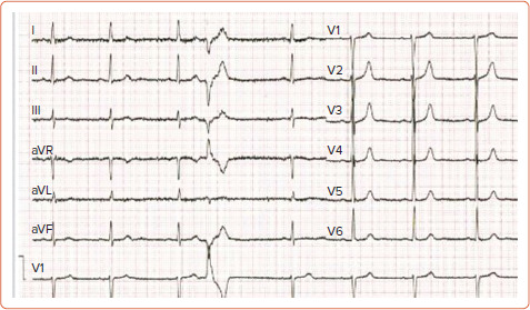
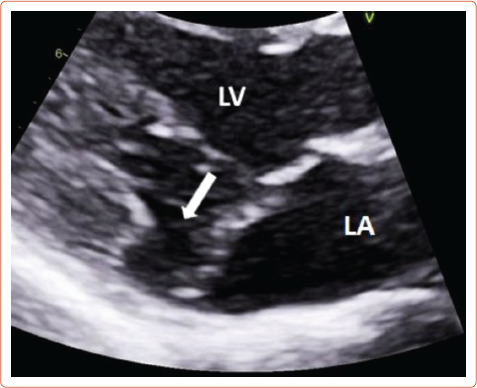
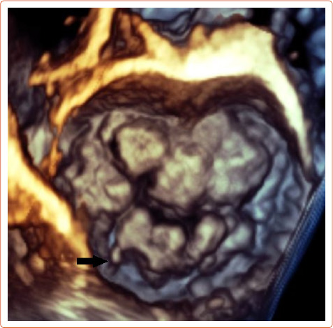
Mitral valve prolapse (MVP) is a common condition present in 1-3% of the population. There has been evidence that a subset of MVP patients is at higher risk of sudden cardiac death. The arrhythmogenic mechanism is related to fibrotic changes in the papillary muscles caused by the prolapsing valve. ECG features include ST-segment depression, T wave inversion or biphasic T waves in inferior leads, and premature ventricular contractions arising from the papillary muscles and the fascicular system. Echocardiography can identify MVP and mitral annular disjunction, a feature that has significant negative prognostic value in MVP. Cardiac MRI is indicated for identifying fibrosis. Patients with high-risk features should be referred for further evaluation. Catheter ablation and mitral valve repair might reduce the risk of malignant arrhythmia. MVP patients with high-risk features and clinically documented ventricular arrhythmia may also be considered for an ICD.

 求助内容:
求助内容: 应助结果提醒方式:


