{"title":"单侧阻生犬的骨骼和牙槽骨尺寸:锥束计算机断层扫描研究。","authors":"Sanjit Kumar Sar, Mandeep Singh, Ankit Sharma, Payal Sharma, Monis Raza","doi":"10.26650/eor.2022911925","DOIUrl":null,"url":null,"abstract":"<p><strong>Purpose: </strong>To compare skeletal and dentoalveolar measurements of subjects with unilateral impacted canine versus the non-impacted contralateral side using cone beam computed tomography (CBCT).</p><p><strong>Materials and methods: </strong>30 CBCTs with unilaterally impacted maxillary canines (Buccal=15, Palatal=15) were selected. Skeletal and dentoalveolar variables (alveolar ridge height of incisors, dentoalveolar height, angulations of incisors and canines, basal lateral width and premolar width) were compared between the impacted and the contralateral sides. Independent t-test was used to compare the variables.</p><p><strong>Results: </strong>There was a significant difference in the mean basal lateral width between the impacted (28.25±1.83 mm) and non-impacted (31.64±2.18 mm) sides. Premolar width was significantly lower on the impacted side (p<0.05). The canines exhibited significantly greater angulations on the impacted side compared to the nonimpacted side. The basal lateral width was significantly higher in the buccal subgroup (29.03±1.65mm) compared to palatal (27.48±1.70mm) on the impacted side. The intra-operator reliability was found to be high (0.99%).</p><p><strong>Conclusion: </strong>Significant differences were seen in canine angulation, premolar width and basal lateral width between impacted vs. non impacted sides. Basal lateral width was higher in buccal impacted cases compared to palatal.</p>","PeriodicalId":41993,"journal":{"name":"European Oral Research","volume":"56 2","pages":"74-79"},"PeriodicalIF":1.1000,"publicationDate":"2022-05-05","publicationTypes":"Journal Article","fieldsOfStudy":null,"isOpenAccess":false,"openAccessPdf":"https://ftp.ncbi.nlm.nih.gov/pub/pmc/oa_pdf/44/b3/eor-056-074.PMC9377773.pdf","citationCount":"2","resultStr":"{\"title\":\"Skeletal and dentoalveolar dimensions in unilateral impacted canines: a cone beam computed tomography study.\",\"authors\":\"Sanjit Kumar Sar, Mandeep Singh, Ankit Sharma, Payal Sharma, Monis Raza\",\"doi\":\"10.26650/eor.2022911925\",\"DOIUrl\":null,\"url\":null,\"abstract\":\"<p><strong>Purpose: </strong>To compare skeletal and dentoalveolar measurements of subjects with unilateral impacted canine versus the non-impacted contralateral side using cone beam computed tomography (CBCT).</p><p><strong>Materials and methods: </strong>30 CBCTs with unilaterally impacted maxillary canines (Buccal=15, Palatal=15) were selected. Skeletal and dentoalveolar variables (alveolar ridge height of incisors, dentoalveolar height, angulations of incisors and canines, basal lateral width and premolar width) were compared between the impacted and the contralateral sides. Independent t-test was used to compare the variables.</p><p><strong>Results: </strong>There was a significant difference in the mean basal lateral width between the impacted (28.25±1.83 mm) and non-impacted (31.64±2.18 mm) sides. Premolar width was significantly lower on the impacted side (p<0.05). The canines exhibited significantly greater angulations on the impacted side compared to the nonimpacted side. The basal lateral width was significantly higher in the buccal subgroup (29.03±1.65mm) compared to palatal (27.48±1.70mm) on the impacted side. The intra-operator reliability was found to be high (0.99%).</p><p><strong>Conclusion: </strong>Significant differences were seen in canine angulation, premolar width and basal lateral width between impacted vs. non impacted sides. Basal lateral width was higher in buccal impacted cases compared to palatal.</p>\",\"PeriodicalId\":41993,\"journal\":{\"name\":\"European Oral Research\",\"volume\":\"56 2\",\"pages\":\"74-79\"},\"PeriodicalIF\":1.1000,\"publicationDate\":\"2022-05-05\",\"publicationTypes\":\"Journal Article\",\"fieldsOfStudy\":null,\"isOpenAccess\":false,\"openAccessPdf\":\"https://ftp.ncbi.nlm.nih.gov/pub/pmc/oa_pdf/44/b3/eor-056-074.PMC9377773.pdf\",\"citationCount\":\"2\",\"resultStr\":null,\"platform\":\"Semanticscholar\",\"paperid\":null,\"PeriodicalName\":\"European Oral Research\",\"FirstCategoryId\":\"1085\",\"ListUrlMain\":\"https://doi.org/10.26650/eor.2022911925\",\"RegionNum\":0,\"RegionCategory\":null,\"ArticlePicture\":[],\"TitleCN\":null,\"AbstractTextCN\":null,\"PMCID\":null,\"EPubDate\":\"\",\"PubModel\":\"\",\"JCR\":\"Q3\",\"JCRName\":\"DENTISTRY, ORAL SURGERY & MEDICINE\",\"Score\":null,\"Total\":0}","platform":"Semanticscholar","paperid":null,"PeriodicalName":"European Oral Research","FirstCategoryId":"1085","ListUrlMain":"https://doi.org/10.26650/eor.2022911925","RegionNum":0,"RegionCategory":null,"ArticlePicture":[],"TitleCN":null,"AbstractTextCN":null,"PMCID":null,"EPubDate":"","PubModel":"","JCR":"Q3","JCRName":"DENTISTRY, ORAL SURGERY & MEDICINE","Score":null,"Total":0}
Skeletal and dentoalveolar dimensions in unilateral impacted canines: a cone beam computed tomography study.
Purpose: To compare skeletal and dentoalveolar measurements of subjects with unilateral impacted canine versus the non-impacted contralateral side using cone beam computed tomography (CBCT).
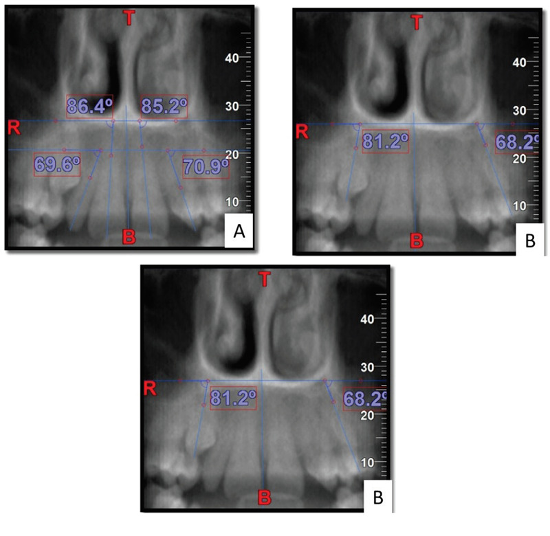
Materials and methods: 30 CBCTs with unilaterally impacted maxillary canines (Buccal=15, Palatal=15) were selected. Skeletal and dentoalveolar variables (alveolar ridge height of incisors, dentoalveolar height, angulations of incisors and canines, basal lateral width and premolar width) were compared between the impacted and the contralateral sides. Independent t-test was used to compare the variables.
Results: There was a significant difference in the mean basal lateral width between the impacted (28.25±1.83 mm) and non-impacted (31.64±2.18 mm) sides. Premolar width was significantly lower on the impacted side (p<0.05). The canines exhibited significantly greater angulations on the impacted side compared to the nonimpacted side. The basal lateral width was significantly higher in the buccal subgroup (29.03±1.65mm) compared to palatal (27.48±1.70mm) on the impacted side. The intra-operator reliability was found to be high (0.99%).
Conclusion: Significant differences were seen in canine angulation, premolar width and basal lateral width between impacted vs. non impacted sides. Basal lateral width was higher in buccal impacted cases compared to palatal.


 求助内容:
求助内容: 应助结果提醒方式:


