{"title":"肉芽肿合并多血管炎1例,多囊性鼻中隔脓肿辅助诊断。","authors":"Mayuko Sasawaki, Kazuhiro Omura, Teru Ebihara, Nobuyoshi Otori","doi":"10.1155/2022/7415498","DOIUrl":null,"url":null,"abstract":"<p><p>A 69-year-old male patient presented to the hospital with a chief complaint of nasal obstruction. Physical examination revealed swelling of the anterior nasal septum and nasal dorsum and tender indurated oedema of the dorsum of both hands. Blood tests showed an elevated inflammatory response, and contrast-enhanced computed tomography (CT) showed a polycystic abscess in the nasal septum. Emergency surgery and histopathology were performed on the day of the initial visit for incisional drainage. Intraoperative findings showed white necrosis between the nasal septal cartilage and nasal septal mucosa, as well as white necrosis and pus accumulation in the periosteum and soft tissue of the piriform aperture and the nasal bone. The patient underwent endoscopic dissection and drained as much as possible, and the abscess and surrounding normal nasal septal mucosa were sampled for diagnostic purposes. The patient was diagnosed with vasculitis based on the clinical findings, pathological examination results, and blood test results. After the diagnosis was confirmed, steroid and cyclophosphamide pulse administration was initiated, and the swelling of the anterior nasal septum and nasal dorsum and the bilateral dorsal indentation oedema improved markedly. The patient is now doing well and will continue to be carefully monitored in the outpatient clinic.</p>","PeriodicalId":45872,"journal":{"name":"Case Reports in Otolaryngology","volume":null,"pages":null},"PeriodicalIF":0.4000,"publicationDate":"2022-10-12","publicationTypes":"Journal Article","fieldsOfStudy":null,"isOpenAccess":false,"openAccessPdf":"https://www.ncbi.nlm.nih.gov/pmc/articles/PMC9581696/pdf/","citationCount":"1","resultStr":"{\"title\":\"A Case of Granulomatosis with Polyangiitis (GPA) Where a Multicystic Nasal Septal Abscess Aided in the Diagnosis.\",\"authors\":\"Mayuko Sasawaki, Kazuhiro Omura, Teru Ebihara, Nobuyoshi Otori\",\"doi\":\"10.1155/2022/7415498\",\"DOIUrl\":null,\"url\":null,\"abstract\":\"<p><p>A 69-year-old male patient presented to the hospital with a chief complaint of nasal obstruction. Physical examination revealed swelling of the anterior nasal septum and nasal dorsum and tender indurated oedema of the dorsum of both hands. Blood tests showed an elevated inflammatory response, and contrast-enhanced computed tomography (CT) showed a polycystic abscess in the nasal septum. Emergency surgery and histopathology were performed on the day of the initial visit for incisional drainage. Intraoperative findings showed white necrosis between the nasal septal cartilage and nasal septal mucosa, as well as white necrosis and pus accumulation in the periosteum and soft tissue of the piriform aperture and the nasal bone. The patient underwent endoscopic dissection and drained as much as possible, and the abscess and surrounding normal nasal septal mucosa were sampled for diagnostic purposes. The patient was diagnosed with vasculitis based on the clinical findings, pathological examination results, and blood test results. After the diagnosis was confirmed, steroid and cyclophosphamide pulse administration was initiated, and the swelling of the anterior nasal septum and nasal dorsum and the bilateral dorsal indentation oedema improved markedly. The patient is now doing well and will continue to be carefully monitored in the outpatient clinic.</p>\",\"PeriodicalId\":45872,\"journal\":{\"name\":\"Case Reports in Otolaryngology\",\"volume\":null,\"pages\":null},\"PeriodicalIF\":0.4000,\"publicationDate\":\"2022-10-12\",\"publicationTypes\":\"Journal Article\",\"fieldsOfStudy\":null,\"isOpenAccess\":false,\"openAccessPdf\":\"https://www.ncbi.nlm.nih.gov/pmc/articles/PMC9581696/pdf/\",\"citationCount\":\"1\",\"resultStr\":null,\"platform\":\"Semanticscholar\",\"paperid\":null,\"PeriodicalName\":\"Case Reports in Otolaryngology\",\"FirstCategoryId\":\"1085\",\"ListUrlMain\":\"https://doi.org/10.1155/2022/7415498\",\"RegionNum\":0,\"RegionCategory\":null,\"ArticlePicture\":[],\"TitleCN\":null,\"AbstractTextCN\":null,\"PMCID\":null,\"EPubDate\":\"2022/1/1 0:00:00\",\"PubModel\":\"eCollection\",\"JCR\":\"Q4\",\"JCRName\":\"OTORHINOLARYNGOLOGY\",\"Score\":null,\"Total\":0}","platform":"Semanticscholar","paperid":null,"PeriodicalName":"Case Reports in Otolaryngology","FirstCategoryId":"1085","ListUrlMain":"https://doi.org/10.1155/2022/7415498","RegionNum":0,"RegionCategory":null,"ArticlePicture":[],"TitleCN":null,"AbstractTextCN":null,"PMCID":null,"EPubDate":"2022/1/1 0:00:00","PubModel":"eCollection","JCR":"Q4","JCRName":"OTORHINOLARYNGOLOGY","Score":null,"Total":0}
A Case of Granulomatosis with Polyangiitis (GPA) Where a Multicystic Nasal Septal Abscess Aided in the Diagnosis.
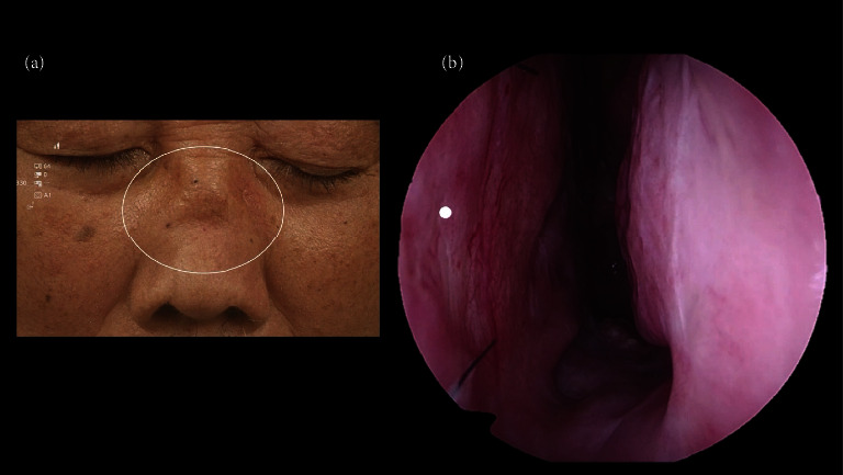
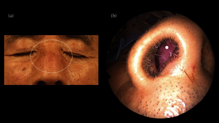
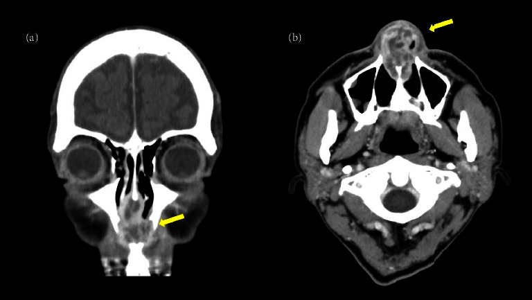
A 69-year-old male patient presented to the hospital with a chief complaint of nasal obstruction. Physical examination revealed swelling of the anterior nasal septum and nasal dorsum and tender indurated oedema of the dorsum of both hands. Blood tests showed an elevated inflammatory response, and contrast-enhanced computed tomography (CT) showed a polycystic abscess in the nasal septum. Emergency surgery and histopathology were performed on the day of the initial visit for incisional drainage. Intraoperative findings showed white necrosis between the nasal septal cartilage and nasal septal mucosa, as well as white necrosis and pus accumulation in the periosteum and soft tissue of the piriform aperture and the nasal bone. The patient underwent endoscopic dissection and drained as much as possible, and the abscess and surrounding normal nasal septal mucosa were sampled for diagnostic purposes. The patient was diagnosed with vasculitis based on the clinical findings, pathological examination results, and blood test results. After the diagnosis was confirmed, steroid and cyclophosphamide pulse administration was initiated, and the swelling of the anterior nasal septum and nasal dorsum and the bilateral dorsal indentation oedema improved markedly. The patient is now doing well and will continue to be carefully monitored in the outpatient clinic.

 求助内容:
求助内容: 应助结果提醒方式:


