{"title":"三甲胺n -氧化物对自发性高血压大鼠内脏白色脂肪组织代谢的影响。","authors":"Guo-Dong He, Xiao-Cong Liu, Xing-Hua Hou, Ying-Qing Feng","doi":"10.1080/21623945.2022.2104783","DOIUrl":null,"url":null,"abstract":"<p><p>Strong links have been reported among trimethylamine N-oxide (TMAO), visceral white adipose tissue (vWAT), and cardiometabolic diseases. However, the effects of TMAO on vWAT in hypertension remained incompletely explored. The impact of a chronic 22-week-long treatment with 1 g/L TMAO on vWAT, and its transcriptional and metabolic changes in spontaneously hypertensive rats (SHRs) were evaluated by serum cytokine measurements, histological analysis, fatty acid determinations, and co-expression network analyses. TMAO increased the serum interleukin-6 levels and insulin secretion in SHRs. The adipocyte size was diminished in the SHR 1 g/L TMAO group. In addition, one kind of monounsaturated fatty acids (cis-15-tetracosenoate) and four kinds of polyunsaturated fatty acids (cis-11,14,17-eicosatrienoic acid, docosatetraenoate, docosapentaenoate n-3, and docosapentaenoate n-6) were elevated by TMAO treatment. Three co-expression modules significantly related to TMAO treatment were identified and pathway enrichment analyses indicated that phagosome, lysosome, fatty acid metabolism, valine, leucine, and isoleucine degradation and metabolic pathways were the most significantly altered biological pathways. This study shed new light on the metabolic roles of TMAO on the vWAT of SHRs. TMAO regulated the metabolic status of vWAT, including reduced lipogenesis and an improved specific fatty acid composition. The mechanisms underlying these effects likely involve phagosome and lysosome pathways.</p>","PeriodicalId":7226,"journal":{"name":"Adipocyte","volume":" ","pages":"420-433"},"PeriodicalIF":3.1000,"publicationDate":"2022-12-01","publicationTypes":"Journal Article","fieldsOfStudy":null,"isOpenAccess":false,"openAccessPdf":"https://www.ncbi.nlm.nih.gov/pmc/articles/PMC9387326/pdf/","citationCount":"1","resultStr":"{\"title\":\"The effect of trimethylamine N-oxide on the metabolism of visceral white adipose tissue in spontaneously hypertensive rat.\",\"authors\":\"Guo-Dong He, Xiao-Cong Liu, Xing-Hua Hou, Ying-Qing Feng\",\"doi\":\"10.1080/21623945.2022.2104783\",\"DOIUrl\":null,\"url\":null,\"abstract\":\"<p><p>Strong links have been reported among trimethylamine N-oxide (TMAO), visceral white adipose tissue (vWAT), and cardiometabolic diseases. However, the effects of TMAO on vWAT in hypertension remained incompletely explored. The impact of a chronic 22-week-long treatment with 1 g/L TMAO on vWAT, and its transcriptional and metabolic changes in spontaneously hypertensive rats (SHRs) were evaluated by serum cytokine measurements, histological analysis, fatty acid determinations, and co-expression network analyses. TMAO increased the serum interleukin-6 levels and insulin secretion in SHRs. The adipocyte size was diminished in the SHR 1 g/L TMAO group. In addition, one kind of monounsaturated fatty acids (cis-15-tetracosenoate) and four kinds of polyunsaturated fatty acids (cis-11,14,17-eicosatrienoic acid, docosatetraenoate, docosapentaenoate n-3, and docosapentaenoate n-6) were elevated by TMAO treatment. Three co-expression modules significantly related to TMAO treatment were identified and pathway enrichment analyses indicated that phagosome, lysosome, fatty acid metabolism, valine, leucine, and isoleucine degradation and metabolic pathways were the most significantly altered biological pathways. This study shed new light on the metabolic roles of TMAO on the vWAT of SHRs. TMAO regulated the metabolic status of vWAT, including reduced lipogenesis and an improved specific fatty acid composition. The mechanisms underlying these effects likely involve phagosome and lysosome pathways.</p>\",\"PeriodicalId\":7226,\"journal\":{\"name\":\"Adipocyte\",\"volume\":\" \",\"pages\":\"420-433\"},\"PeriodicalIF\":3.1000,\"publicationDate\":\"2022-12-01\",\"publicationTypes\":\"Journal Article\",\"fieldsOfStudy\":null,\"isOpenAccess\":false,\"openAccessPdf\":\"https://www.ncbi.nlm.nih.gov/pmc/articles/PMC9387326/pdf/\",\"citationCount\":\"1\",\"resultStr\":null,\"platform\":\"Semanticscholar\",\"paperid\":null,\"PeriodicalName\":\"Adipocyte\",\"FirstCategoryId\":\"99\",\"ListUrlMain\":\"https://doi.org/10.1080/21623945.2022.2104783\",\"RegionNum\":4,\"RegionCategory\":\"生物学\",\"ArticlePicture\":[],\"TitleCN\":null,\"AbstractTextCN\":null,\"PMCID\":null,\"EPubDate\":\"\",\"PubModel\":\"\",\"JCR\":\"Q2\",\"JCRName\":\"ENDOCRINOLOGY & METABOLISM\",\"Score\":null,\"Total\":0}","platform":"Semanticscholar","paperid":null,"PeriodicalName":"Adipocyte","FirstCategoryId":"99","ListUrlMain":"https://doi.org/10.1080/21623945.2022.2104783","RegionNum":4,"RegionCategory":"生物学","ArticlePicture":[],"TitleCN":null,"AbstractTextCN":null,"PMCID":null,"EPubDate":"","PubModel":"","JCR":"Q2","JCRName":"ENDOCRINOLOGY & METABOLISM","Score":null,"Total":0}
The effect of trimethylamine N-oxide on the metabolism of visceral white adipose tissue in spontaneously hypertensive rat.
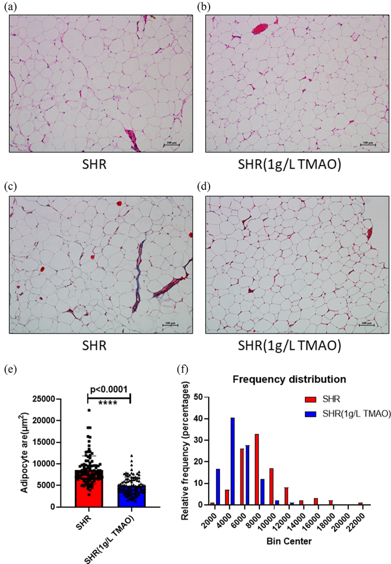
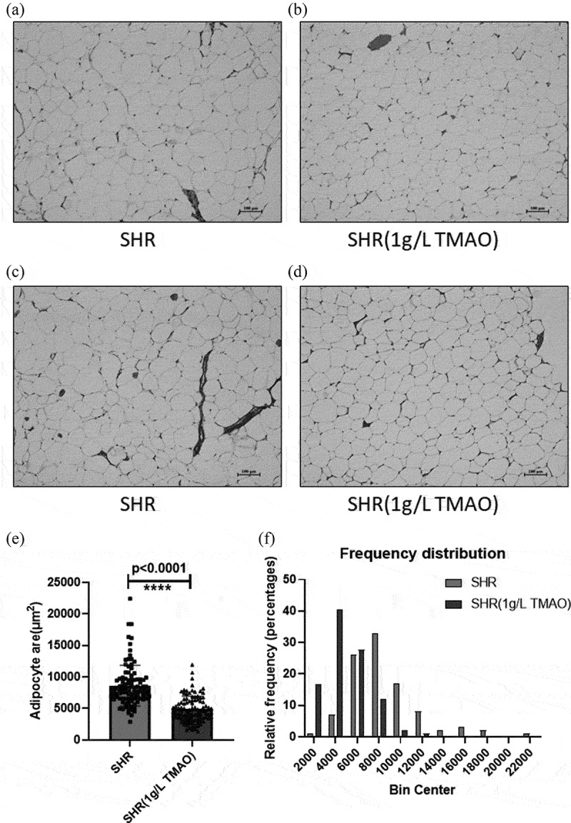
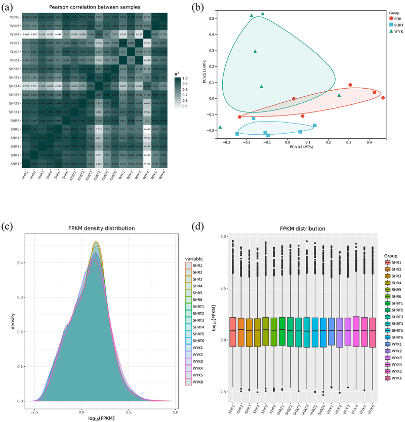
Strong links have been reported among trimethylamine N-oxide (TMAO), visceral white adipose tissue (vWAT), and cardiometabolic diseases. However, the effects of TMAO on vWAT in hypertension remained incompletely explored. The impact of a chronic 22-week-long treatment with 1 g/L TMAO on vWAT, and its transcriptional and metabolic changes in spontaneously hypertensive rats (SHRs) were evaluated by serum cytokine measurements, histological analysis, fatty acid determinations, and co-expression network analyses. TMAO increased the serum interleukin-6 levels and insulin secretion in SHRs. The adipocyte size was diminished in the SHR 1 g/L TMAO group. In addition, one kind of monounsaturated fatty acids (cis-15-tetracosenoate) and four kinds of polyunsaturated fatty acids (cis-11,14,17-eicosatrienoic acid, docosatetraenoate, docosapentaenoate n-3, and docosapentaenoate n-6) were elevated by TMAO treatment. Three co-expression modules significantly related to TMAO treatment were identified and pathway enrichment analyses indicated that phagosome, lysosome, fatty acid metabolism, valine, leucine, and isoleucine degradation and metabolic pathways were the most significantly altered biological pathways. This study shed new light on the metabolic roles of TMAO on the vWAT of SHRs. TMAO regulated the metabolic status of vWAT, including reduced lipogenesis and an improved specific fatty acid composition. The mechanisms underlying these effects likely involve phagosome and lysosome pathways.
期刊介绍:
Adipocyte recognizes that the adipose tissue is the largest endocrine organ in the body, and explores the link between dysfunctional adipose tissue and the growing number of chronic diseases including diabetes, hypertension, cardiovascular disease and cancer. Historically, the primary function of the adipose tissue was limited to energy storage and thermoregulation. However, a plethora of research over the past 3 decades has recognized the dynamic role of the adipose tissue and its contribution to a variety of physiological processes including reproduction, angiogenesis, apoptosis, inflammation, blood pressure, coagulation, fibrinolysis, immunity and general metabolic homeostasis. The field of Adipose Tissue research has grown tremendously, and Adipocyte is the first international peer-reviewed journal of its kind providing a multi-disciplinary forum for research focusing exclusively on all aspects of adipose tissue physiology and pathophysiology. Adipocyte accepts high-profile submissions in basic, translational and clinical research.
 求助内容:
求助内容: 应助结果提醒方式:


