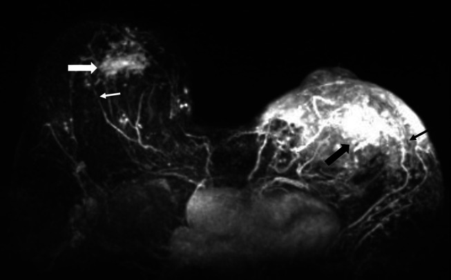
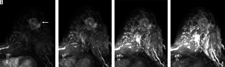
{"title":"乳腺恶性肿瘤中邻近血管征象的价值。","authors":"Ezra Çetinkaya, Şeyma Yıldız, Hafize Otçu, Rasul Sharifov, Fatma Çelik Yabul, Alpay Alkan","doi":"10.5152/dir.2022.211228","DOIUrl":null,"url":null,"abstract":"<p><p>PURPOSE The aim of this study was to evaluate the prognostic quality of adjacent vessel sign (AVS) in malignant breast tumors by comparing it with classical prognostic pathological biomarkers and magnetic resonance imaging (MRI) findings. METHODS A total of 124 patients with 133 malignant lesions were included. All the imaging was performed on a 1.5T Avanto scanner and the images were interpreted according to BI-RADS-MR® (fifth ed.) atlas. Maximum intensity projection (MIP) images were constructed from subtracted post-contrast images and were used to investigate AVS. Histopathological results and MRI findings were compared with AVS. RESULTS Interobserver agreement about AVS status was substantial (κ=0.64). AVS positive lesions were significantly bigger in size (P < .001, AVS negative: median 12 mm, AVS positive: median 31 mm). AVS was significantly associated with increased Ki-67 index and axillary lymph node metastasis (P=.009 and P=.019, respectively). Between AVS and lymphovascular invasion (LVI), there was a trend toward positive relationship (P=.076). MRI findings of T2 hypointensity, peritumoral edema, irregular shape, non-homogeneous contrast enhancement, rapid early contrast enhancement, and skin infiltration showed significant positive relation with AVS (P < .001, P < .001, P < .001, P=.02, P=.021, and P=.021, respectively). AVS is found to be associated with increased Ki-67 index, axillary lymph node metastasis, and some MRI findings that point to malignancy or poor prognosis. CONCLUSION AVS indicates poor prognosis since it is related to axillary lymph node metastasis, increased Ki-67 index, LVI, peritumoral edema, rapid early contrast enhancement, increased background enhancement, skin extension, T2 hypointensity, non-homogeneous contrast enhancement, irregular lesion shape, and larger tumor size. AVS is an easy to use sign that shows substantial interobserver agreement.</p>","PeriodicalId":50582,"journal":{"name":"Diagnostic and Interventional Radiology","volume":" ","pages":"463-469"},"PeriodicalIF":1.7000,"publicationDate":"2022-09-01","publicationTypes":"Journal Article","fieldsOfStudy":null,"isOpenAccess":false,"openAccessPdf":"https://ftp.ncbi.nlm.nih.gov/pub/pmc/oa_pdf/7b/08/dir-28-5-463.PMC9682586.pdf","citationCount":"0","resultStr":"{\"title\":\"The value of adjacent vessel sign in malignant breast tumors.\",\"authors\":\"Ezra Çetinkaya, Şeyma Yıldız, Hafize Otçu, Rasul Sharifov, Fatma Çelik Yabul, Alpay Alkan\",\"doi\":\"10.5152/dir.2022.211228\",\"DOIUrl\":null,\"url\":null,\"abstract\":\"<p><p>PURPOSE The aim of this study was to evaluate the prognostic quality of adjacent vessel sign (AVS) in malignant breast tumors by comparing it with classical prognostic pathological biomarkers and magnetic resonance imaging (MRI) findings. METHODS A total of 124 patients with 133 malignant lesions were included. All the imaging was performed on a 1.5T Avanto scanner and the images were interpreted according to BI-RADS-MR® (fifth ed.) atlas. Maximum intensity projection (MIP) images were constructed from subtracted post-contrast images and were used to investigate AVS. Histopathological results and MRI findings were compared with AVS. RESULTS Interobserver agreement about AVS status was substantial (κ=0.64). AVS positive lesions were significantly bigger in size (P < .001, AVS negative: median 12 mm, AVS positive: median 31 mm). AVS was significantly associated with increased Ki-67 index and axillary lymph node metastasis (P=.009 and P=.019, respectively). Between AVS and lymphovascular invasion (LVI), there was a trend toward positive relationship (P=.076). MRI findings of T2 hypointensity, peritumoral edema, irregular shape, non-homogeneous contrast enhancement, rapid early contrast enhancement, and skin infiltration showed significant positive relation with AVS (P < .001, P < .001, P < .001, P=.02, P=.021, and P=.021, respectively). AVS is found to be associated with increased Ki-67 index, axillary lymph node metastasis, and some MRI findings that point to malignancy or poor prognosis. CONCLUSION AVS indicates poor prognosis since it is related to axillary lymph node metastasis, increased Ki-67 index, LVI, peritumoral edema, rapid early contrast enhancement, increased background enhancement, skin extension, T2 hypointensity, non-homogeneous contrast enhancement, irregular lesion shape, and larger tumor size. AVS is an easy to use sign that shows substantial interobserver agreement.</p>\",\"PeriodicalId\":50582,\"journal\":{\"name\":\"Diagnostic and Interventional Radiology\",\"volume\":\" \",\"pages\":\"463-469\"},\"PeriodicalIF\":1.7000,\"publicationDate\":\"2022-09-01\",\"publicationTypes\":\"Journal Article\",\"fieldsOfStudy\":null,\"isOpenAccess\":false,\"openAccessPdf\":\"https://ftp.ncbi.nlm.nih.gov/pub/pmc/oa_pdf/7b/08/dir-28-5-463.PMC9682586.pdf\",\"citationCount\":\"0\",\"resultStr\":null,\"platform\":\"Semanticscholar\",\"paperid\":null,\"PeriodicalName\":\"Diagnostic and Interventional Radiology\",\"FirstCategoryId\":\"3\",\"ListUrlMain\":\"https://doi.org/10.5152/dir.2022.211228\",\"RegionNum\":4,\"RegionCategory\":\"医学\",\"ArticlePicture\":[],\"TitleCN\":null,\"AbstractTextCN\":null,\"PMCID\":null,\"EPubDate\":\"\",\"PubModel\":\"\",\"JCR\":\"Q2\",\"JCRName\":\"Medicine\",\"Score\":null,\"Total\":0}","platform":"Semanticscholar","paperid":null,"PeriodicalName":"Diagnostic and Interventional Radiology","FirstCategoryId":"3","ListUrlMain":"https://doi.org/10.5152/dir.2022.211228","RegionNum":4,"RegionCategory":"医学","ArticlePicture":[],"TitleCN":null,"AbstractTextCN":null,"PMCID":null,"EPubDate":"","PubModel":"","JCR":"Q2","JCRName":"Medicine","Score":null,"Total":0}
引用次数: 0
摘要
目的通过与经典预后病理生物标志物和磁共振成像(MRI)结果的比较,评价乳腺恶性肿瘤中邻近血管征象(AVS)的预后质量。方法回顾性分析124例恶性肿瘤133例。所有成像均在1.5T Avanto扫描仪上进行,图像根据BI-RADS-MR®(第五版)图谱进行解释。最大强度投影(MIP)图像由减相后图像构建,用于研究AVS。将组织病理学结果与MRI结果与AVS进行比较。结果观察间AVS状态的一致性显著(κ=0.64)。AVS阳性病变体积显著增大(P < 0.001, AVS阴性:中位12 mm, AVS阳性:中位31 mm)。AVS与Ki-67指数升高及腋窝淋巴结转移显著相关(P=。009, P=。019年,分别)。AVS与淋巴血管侵袭(LVI)呈显著正相关(P= 0.076)。T2低信号、瘤周水肿、形状不规则、增强不均匀、早期快速增强、皮肤浸润等MRI表现与AVS呈显著正相关(P < 0.001, P < 0.001, P < 0.001, P=)。02年,P =。021, P=。021年,分别)。AVS与Ki-67指数升高、腋窝淋巴结转移以及一些提示恶性或预后不良的MRI表现相关。结论AVS预后较差,与腋窝淋巴结转移、Ki-67指数升高、LVI、瘤周水肿、早期增强快速、背景增强增加、皮肤延伸、T2低密度、增强不均匀、病变形状不规则、肿瘤体积较大有关。AVS是一种易于使用的符号,显示了大量的观察者之间的协议。
The value of adjacent vessel sign in malignant breast tumors.
PURPOSE The aim of this study was to evaluate the prognostic quality of adjacent vessel sign (AVS) in malignant breast tumors by comparing it with classical prognostic pathological biomarkers and magnetic resonance imaging (MRI) findings. METHODS A total of 124 patients with 133 malignant lesions were included. All the imaging was performed on a 1.5T Avanto scanner and the images were interpreted according to BI-RADS-MR® (fifth ed.) atlas. Maximum intensity projection (MIP) images were constructed from subtracted post-contrast images and were used to investigate AVS. Histopathological results and MRI findings were compared with AVS. RESULTS Interobserver agreement about AVS status was substantial (κ=0.64). AVS positive lesions were significantly bigger in size (P < .001, AVS negative: median 12 mm, AVS positive: median 31 mm). AVS was significantly associated with increased Ki-67 index and axillary lymph node metastasis (P=.009 and P=.019, respectively). Between AVS and lymphovascular invasion (LVI), there was a trend toward positive relationship (P=.076). MRI findings of T2 hypointensity, peritumoral edema, irregular shape, non-homogeneous contrast enhancement, rapid early contrast enhancement, and skin infiltration showed significant positive relation with AVS (P < .001, P < .001, P < .001, P=.02, P=.021, and P=.021, respectively). AVS is found to be associated with increased Ki-67 index, axillary lymph node metastasis, and some MRI findings that point to malignancy or poor prognosis. CONCLUSION AVS indicates poor prognosis since it is related to axillary lymph node metastasis, increased Ki-67 index, LVI, peritumoral edema, rapid early contrast enhancement, increased background enhancement, skin extension, T2 hypointensity, non-homogeneous contrast enhancement, irregular lesion shape, and larger tumor size. AVS is an easy to use sign that shows substantial interobserver agreement.
期刊介绍:
Diagnostic and Interventional Radiology (Diagn Interv Radiol) is the open access, online-only official publication of Turkish Society of Radiology. It is published bimonthly and the journal’s publication language is English.
The journal is a medium for original articles, reviews, pictorial essays, technical notes related to all fields of diagnostic and interventional radiology.


 求助内容:
求助内容: 应助结果提醒方式:


