{"title":"HIV免疫抑制患者的疱疹病毒假瘤。","authors":"Daniel York, Pavan Patel, Smera Saikumar","doi":"10.1155/2022/3109331","DOIUrl":null,"url":null,"abstract":"<p><strong>Background: </strong>In rare cases, HSV infections can present as pseudotumors that are often mistaken as malignancies in patients with an uncontrolled HIV infection. Herpes simplex virus type 2 (HSV-2) infection rates range from 60% to 90% in individuals coinfected with HIV. <i>Case Presentation</i>. A 48-year-old patient presented with a large fungating mass near her right inferior vulva with a hardness of surrounding tissues. The mass was 4 cm × 3 cm in size and was excised in the operating room. The pathology was negative for malignancy; however, it showed lymphoplasmacytic proliferation with immunostaining positive for HSV virus.</p><p><strong>Conclusion: </strong>Atypical HSV pseudotumors should be considered in the differential diagnosis for an immunosuppressed patient who presents with a genital mass lesion.</p>","PeriodicalId":9610,"journal":{"name":"Case Reports in Obstetrics and Gynecology","volume":" ","pages":"3109331"},"PeriodicalIF":0.8000,"publicationDate":"2022-07-07","publicationTypes":"Journal Article","fieldsOfStudy":null,"isOpenAccess":false,"openAccessPdf":"https://www.ncbi.nlm.nih.gov/pmc/articles/PMC9283043/pdf/","citationCount":"0","resultStr":"{\"title\":\"Herpes Virus Pseudotumor in a Patient with HIV Immunosuppression.\",\"authors\":\"Daniel York, Pavan Patel, Smera Saikumar\",\"doi\":\"10.1155/2022/3109331\",\"DOIUrl\":null,\"url\":null,\"abstract\":\"<p><strong>Background: </strong>In rare cases, HSV infections can present as pseudotumors that are often mistaken as malignancies in patients with an uncontrolled HIV infection. Herpes simplex virus type 2 (HSV-2) infection rates range from 60% to 90% in individuals coinfected with HIV. <i>Case Presentation</i>. A 48-year-old patient presented with a large fungating mass near her right inferior vulva with a hardness of surrounding tissues. The mass was 4 cm × 3 cm in size and was excised in the operating room. The pathology was negative for malignancy; however, it showed lymphoplasmacytic proliferation with immunostaining positive for HSV virus.</p><p><strong>Conclusion: </strong>Atypical HSV pseudotumors should be considered in the differential diagnosis for an immunosuppressed patient who presents with a genital mass lesion.</p>\",\"PeriodicalId\":9610,\"journal\":{\"name\":\"Case Reports in Obstetrics and Gynecology\",\"volume\":\" \",\"pages\":\"3109331\"},\"PeriodicalIF\":0.8000,\"publicationDate\":\"2022-07-07\",\"publicationTypes\":\"Journal Article\",\"fieldsOfStudy\":null,\"isOpenAccess\":false,\"openAccessPdf\":\"https://www.ncbi.nlm.nih.gov/pmc/articles/PMC9283043/pdf/\",\"citationCount\":\"0\",\"resultStr\":null,\"platform\":\"Semanticscholar\",\"paperid\":null,\"PeriodicalName\":\"Case Reports in Obstetrics and Gynecology\",\"FirstCategoryId\":\"1085\",\"ListUrlMain\":\"https://doi.org/10.1155/2022/3109331\",\"RegionNum\":0,\"RegionCategory\":null,\"ArticlePicture\":[],\"TitleCN\":null,\"AbstractTextCN\":null,\"PMCID\":null,\"EPubDate\":\"2022/1/1 0:00:00\",\"PubModel\":\"eCollection\",\"JCR\":\"Q4\",\"JCRName\":\"OBSTETRICS & GYNECOLOGY\",\"Score\":null,\"Total\":0}","platform":"Semanticscholar","paperid":null,"PeriodicalName":"Case Reports in Obstetrics and Gynecology","FirstCategoryId":"1085","ListUrlMain":"https://doi.org/10.1155/2022/3109331","RegionNum":0,"RegionCategory":null,"ArticlePicture":[],"TitleCN":null,"AbstractTextCN":null,"PMCID":null,"EPubDate":"2022/1/1 0:00:00","PubModel":"eCollection","JCR":"Q4","JCRName":"OBSTETRICS & GYNECOLOGY","Score":null,"Total":0}
Herpes Virus Pseudotumor in a Patient with HIV Immunosuppression.
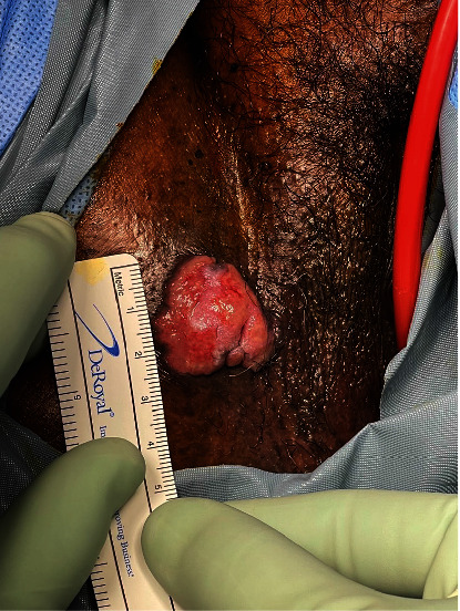
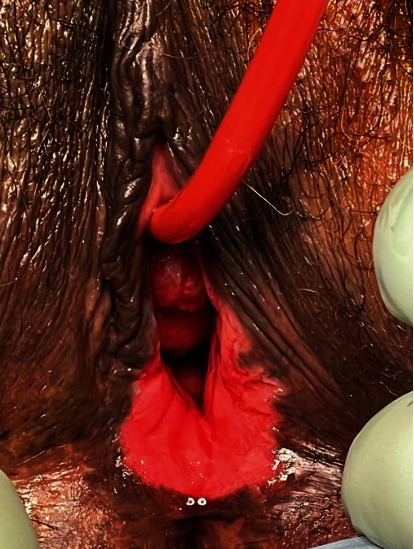
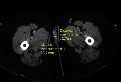
Background: In rare cases, HSV infections can present as pseudotumors that are often mistaken as malignancies in patients with an uncontrolled HIV infection. Herpes simplex virus type 2 (HSV-2) infection rates range from 60% to 90% in individuals coinfected with HIV. Case Presentation. A 48-year-old patient presented with a large fungating mass near her right inferior vulva with a hardness of surrounding tissues. The mass was 4 cm × 3 cm in size and was excised in the operating room. The pathology was negative for malignancy; however, it showed lymphoplasmacytic proliferation with immunostaining positive for HSV virus.
Conclusion: Atypical HSV pseudotumors should be considered in the differential diagnosis for an immunosuppressed patient who presents with a genital mass lesion.

 求助内容:
求助内容: 应助结果提醒方式:


