Gabriella Melson, Elie Saliba, Shreya Patel, Richard Eisen, Candice E Brem
{"title":"具有恶性细胞学特征的透明细胞棘瘤1例报告及文献复习。","authors":"Gabriella Melson, Elie Saliba, Shreya Patel, Richard Eisen, Candice E Brem","doi":"10.3390/dermatopathology9040041","DOIUrl":null,"url":null,"abstract":"<p><p>Clear cell acanthoma (CCA) is classically considered a benign epidermal tumor, although rare case reports have described CCA with malignant features. Here, we present a case of a patient with a biopsy proven CCA that regrew post-biopsy and was subsequently completely excised. Histologic examination of the tumor in the excision specimen revealed malignant cytologic features that were not present in the initial biopsy. A review of the literature identified five additional cases of CCA with similar malignant cytologic features. On analysis, common histopathologic characteristics included cellular pleomorphism, increased nuclear-to-cytoplasmic ratio, prominent nucleoli, and atypical mitotic figures. We support the designation of atypical clear cell acanthoma for these entities with features of both CCA and significant cytologic atypia. As none of these cases exhibited clinically aggressive behavior, further study is warranted.</p>","PeriodicalId":42885,"journal":{"name":"Dermatopathology","volume":" ","pages":"355-360"},"PeriodicalIF":1.7000,"publicationDate":"2022-10-20","publicationTypes":"Journal Article","fieldsOfStudy":null,"isOpenAccess":false,"openAccessPdf":"https://www.ncbi.nlm.nih.gov/pmc/articles/PMC9590090/pdf/","citationCount":"1","resultStr":"{\"title\":\"Clear Cell Acanthoma with Malignant Cytologic Features: A Case Report and Review of the Literature.\",\"authors\":\"Gabriella Melson, Elie Saliba, Shreya Patel, Richard Eisen, Candice E Brem\",\"doi\":\"10.3390/dermatopathology9040041\",\"DOIUrl\":null,\"url\":null,\"abstract\":\"<p><p>Clear cell acanthoma (CCA) is classically considered a benign epidermal tumor, although rare case reports have described CCA with malignant features. Here, we present a case of a patient with a biopsy proven CCA that regrew post-biopsy and was subsequently completely excised. Histologic examination of the tumor in the excision specimen revealed malignant cytologic features that were not present in the initial biopsy. A review of the literature identified five additional cases of CCA with similar malignant cytologic features. On analysis, common histopathologic characteristics included cellular pleomorphism, increased nuclear-to-cytoplasmic ratio, prominent nucleoli, and atypical mitotic figures. We support the designation of atypical clear cell acanthoma for these entities with features of both CCA and significant cytologic atypia. As none of these cases exhibited clinically aggressive behavior, further study is warranted.</p>\",\"PeriodicalId\":42885,\"journal\":{\"name\":\"Dermatopathology\",\"volume\":\" \",\"pages\":\"355-360\"},\"PeriodicalIF\":1.7000,\"publicationDate\":\"2022-10-20\",\"publicationTypes\":\"Journal Article\",\"fieldsOfStudy\":null,\"isOpenAccess\":false,\"openAccessPdf\":\"https://www.ncbi.nlm.nih.gov/pmc/articles/PMC9590090/pdf/\",\"citationCount\":\"1\",\"resultStr\":null,\"platform\":\"Semanticscholar\",\"paperid\":null,\"PeriodicalName\":\"Dermatopathology\",\"FirstCategoryId\":\"1085\",\"ListUrlMain\":\"https://doi.org/10.3390/dermatopathology9040041\",\"RegionNum\":0,\"RegionCategory\":null,\"ArticlePicture\":[],\"TitleCN\":null,\"AbstractTextCN\":null,\"PMCID\":null,\"EPubDate\":\"\",\"PubModel\":\"\",\"JCR\":\"Q3\",\"JCRName\":\"DERMATOLOGY\",\"Score\":null,\"Total\":0}","platform":"Semanticscholar","paperid":null,"PeriodicalName":"Dermatopathology","FirstCategoryId":"1085","ListUrlMain":"https://doi.org/10.3390/dermatopathology9040041","RegionNum":0,"RegionCategory":null,"ArticlePicture":[],"TitleCN":null,"AbstractTextCN":null,"PMCID":null,"EPubDate":"","PubModel":"","JCR":"Q3","JCRName":"DERMATOLOGY","Score":null,"Total":0}
Clear Cell Acanthoma with Malignant Cytologic Features: A Case Report and Review of the Literature.
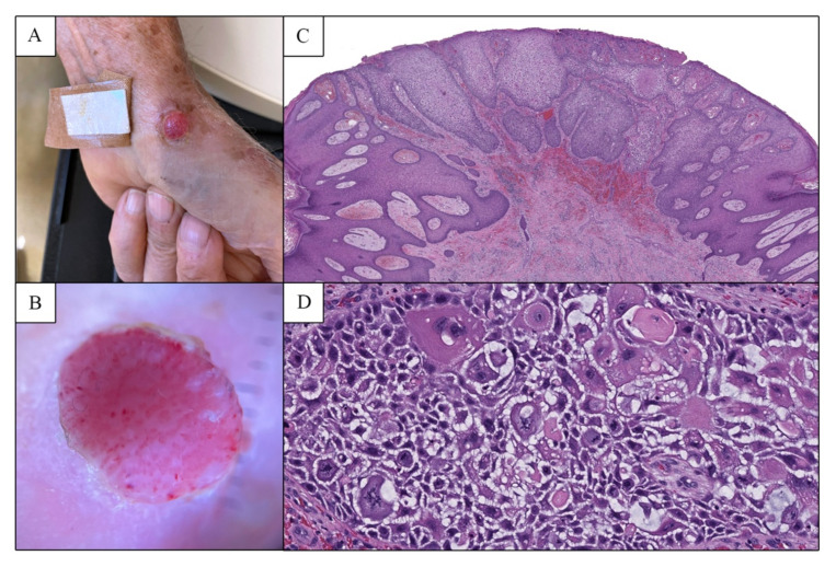
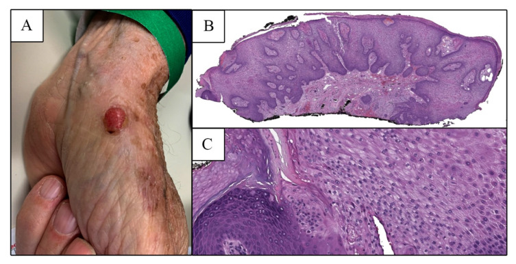
Clear cell acanthoma (CCA) is classically considered a benign epidermal tumor, although rare case reports have described CCA with malignant features. Here, we present a case of a patient with a biopsy proven CCA that regrew post-biopsy and was subsequently completely excised. Histologic examination of the tumor in the excision specimen revealed malignant cytologic features that were not present in the initial biopsy. A review of the literature identified five additional cases of CCA with similar malignant cytologic features. On analysis, common histopathologic characteristics included cellular pleomorphism, increased nuclear-to-cytoplasmic ratio, prominent nucleoli, and atypical mitotic figures. We support the designation of atypical clear cell acanthoma for these entities with features of both CCA and significant cytologic atypia. As none of these cases exhibited clinically aggressive behavior, further study is warranted.

 求助内容:
求助内容: 应助结果提醒方式:


