Antonio Córdoba-Fernández, María Dolores Jiménez-Cristino, Victoria Eugenia Córdoba-Jiménez
{"title":"大小叶毛细血管瘤合并趾甲内生:组织病理学特征及病例报告。","authors":"Antonio Córdoba-Fernández, María Dolores Jiménez-Cristino, Victoria Eugenia Córdoba-Jiménez","doi":"10.3390/dermatopathology9030031","DOIUrl":null,"url":null,"abstract":"<p><p>Lobular capillary hemangioma (LCH-PG) is a type of pyogenic granuloma characterized by proliferating blood vessels that resemble conventional granulation tissue. Granulation tissue is very often seen in association with ingrown toenails. Despite the close relationship between both entities, LCH-PG shows clinically different behaviors, such as rapid growth and frequent recurrence. Currently, it is unknown exactly how the different etiological factors contribute to the formation of differences between entities. We present a case of a large LCH-PG associated with chronic onychocryptosis in a 26-year-old man. Histopathological features included extensive signs of ulceration, hyperkeratosis, and patchy epidermal acanthosis with the presence of fibrous septa with lobular areas beneath the ulcerative area. The presence of stroma with a marked proliferation of blood vessels with wall thickening and mixed-type inflammatory changes was also characteristic. In advanced stages of onychocryptosis, as presented here, conventional granulation tissue or pyogenic granuloma can be clinically difficult to distinguish from other benign or malignant neoplasms. Histological examination is mandatory, and excisional biopsy can provide a definitive diagnosis.</p>","PeriodicalId":42885,"journal":{"name":"Dermatopathology","volume":" ","pages":"271-276"},"PeriodicalIF":1.7000,"publicationDate":"2022-07-25","publicationTypes":"Journal Article","fieldsOfStudy":null,"isOpenAccess":false,"openAccessPdf":"https://www.ncbi.nlm.nih.gov/pmc/articles/PMC9330171/pdf/","citationCount":"0","resultStr":"{\"title\":\"Large Lobular Capillary Hemangioma Associated with Ingrown Toenail: Histopathological Features and Case Report.\",\"authors\":\"Antonio Córdoba-Fernández, María Dolores Jiménez-Cristino, Victoria Eugenia Córdoba-Jiménez\",\"doi\":\"10.3390/dermatopathology9030031\",\"DOIUrl\":null,\"url\":null,\"abstract\":\"<p><p>Lobular capillary hemangioma (LCH-PG) is a type of pyogenic granuloma characterized by proliferating blood vessels that resemble conventional granulation tissue. Granulation tissue is very often seen in association with ingrown toenails. Despite the close relationship between both entities, LCH-PG shows clinically different behaviors, such as rapid growth and frequent recurrence. Currently, it is unknown exactly how the different etiological factors contribute to the formation of differences between entities. We present a case of a large LCH-PG associated with chronic onychocryptosis in a 26-year-old man. Histopathological features included extensive signs of ulceration, hyperkeratosis, and patchy epidermal acanthosis with the presence of fibrous septa with lobular areas beneath the ulcerative area. The presence of stroma with a marked proliferation of blood vessels with wall thickening and mixed-type inflammatory changes was also characteristic. In advanced stages of onychocryptosis, as presented here, conventional granulation tissue or pyogenic granuloma can be clinically difficult to distinguish from other benign or malignant neoplasms. Histological examination is mandatory, and excisional biopsy can provide a definitive diagnosis.</p>\",\"PeriodicalId\":42885,\"journal\":{\"name\":\"Dermatopathology\",\"volume\":\" \",\"pages\":\"271-276\"},\"PeriodicalIF\":1.7000,\"publicationDate\":\"2022-07-25\",\"publicationTypes\":\"Journal Article\",\"fieldsOfStudy\":null,\"isOpenAccess\":false,\"openAccessPdf\":\"https://www.ncbi.nlm.nih.gov/pmc/articles/PMC9330171/pdf/\",\"citationCount\":\"0\",\"resultStr\":null,\"platform\":\"Semanticscholar\",\"paperid\":null,\"PeriodicalName\":\"Dermatopathology\",\"FirstCategoryId\":\"1085\",\"ListUrlMain\":\"https://doi.org/10.3390/dermatopathology9030031\",\"RegionNum\":0,\"RegionCategory\":null,\"ArticlePicture\":[],\"TitleCN\":null,\"AbstractTextCN\":null,\"PMCID\":null,\"EPubDate\":\"\",\"PubModel\":\"\",\"JCR\":\"Q3\",\"JCRName\":\"DERMATOLOGY\",\"Score\":null,\"Total\":0}","platform":"Semanticscholar","paperid":null,"PeriodicalName":"Dermatopathology","FirstCategoryId":"1085","ListUrlMain":"https://doi.org/10.3390/dermatopathology9030031","RegionNum":0,"RegionCategory":null,"ArticlePicture":[],"TitleCN":null,"AbstractTextCN":null,"PMCID":null,"EPubDate":"","PubModel":"","JCR":"Q3","JCRName":"DERMATOLOGY","Score":null,"Total":0}
Large Lobular Capillary Hemangioma Associated with Ingrown Toenail: Histopathological Features and Case Report.
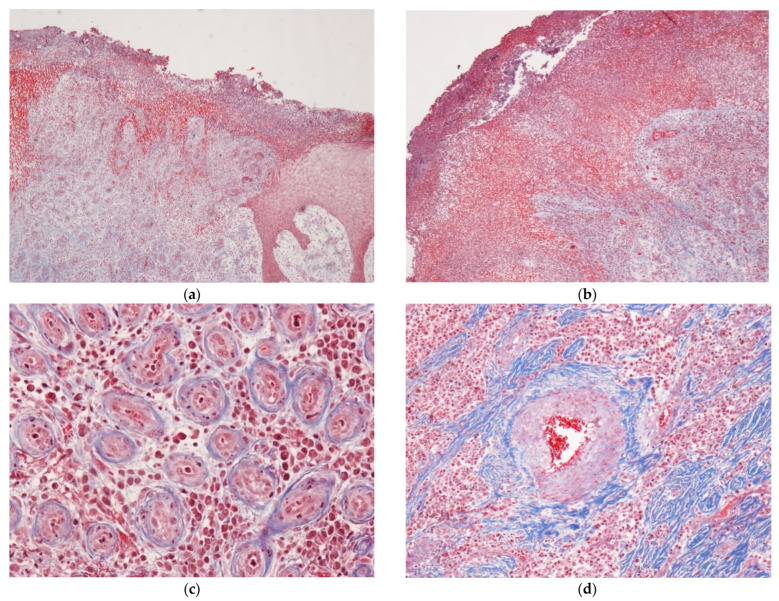
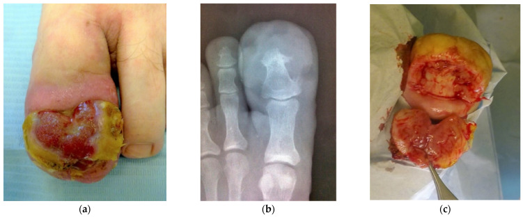
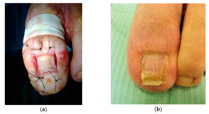
Lobular capillary hemangioma (LCH-PG) is a type of pyogenic granuloma characterized by proliferating blood vessels that resemble conventional granulation tissue. Granulation tissue is very often seen in association with ingrown toenails. Despite the close relationship between both entities, LCH-PG shows clinically different behaviors, such as rapid growth and frequent recurrence. Currently, it is unknown exactly how the different etiological factors contribute to the formation of differences between entities. We present a case of a large LCH-PG associated with chronic onychocryptosis in a 26-year-old man. Histopathological features included extensive signs of ulceration, hyperkeratosis, and patchy epidermal acanthosis with the presence of fibrous septa with lobular areas beneath the ulcerative area. The presence of stroma with a marked proliferation of blood vessels with wall thickening and mixed-type inflammatory changes was also characteristic. In advanced stages of onychocryptosis, as presented here, conventional granulation tissue or pyogenic granuloma can be clinically difficult to distinguish from other benign or malignant neoplasms. Histological examination is mandatory, and excisional biopsy can provide a definitive diagnosis.

 求助内容:
求助内容: 应助结果提醒方式:


