Gabriele Molteni, Michael Ghirelli, Andrea Sacchetto, Matteo Fermi, Stefano De Rossi, Francesco Mattioli, Livio Presutti, Daniele Marchioni
{"title":"使用离体模型的显微外科训练:显微镜与3D外窥镜。","authors":"Gabriele Molteni, Michael Ghirelli, Andrea Sacchetto, Matteo Fermi, Stefano De Rossi, Francesco Mattioli, Livio Presutti, Daniele Marchioni","doi":"10.14639/0392-100X-N1946","DOIUrl":null,"url":null,"abstract":"<p><strong>Objective: </strong>The aim of this study is to evaluate the feasibility of the 3D exoscope in a microvascular anastomosis training setting and compare it with the gold-standard technique using the operating microscope (OM).</p><p><strong>Methods: </strong>Participants were recruited among otorhinolaryngology head and neck surgery (OHNS) residents of two tertiary care hospitals. Trainees were asked to complete 4 microvascular end-to-end anastomoses on chicken thighs with the OM and VITOM 3D exoscope. The performances were scored by experienced microvascular surgeons; an objective evaluation of the anastomosis and a subjective assessment of the workload were conducted.</p><p><strong>Results: </strong>8 OHNS residents were recruited. Considering the amount of time needed to complete (TTC) the anastomosis, an improvement was shown by all the participants throughout the training program. The objective evaluation of the anastomosis did not show a significant difference. No significant differences were found by analyzing the subjective workload with the different tools.</p><p><strong>Conclusions: </strong>This article represents the first attempt to compare the use of the OM and the 3D exoscope during training for microsurgery. The results of our study demonstrate the noninferiority of microsurgical training obtained using the 3D exoscope compared to that offered by the OM.</p>","PeriodicalId":520544,"journal":{"name":"Acta otorhinolaryngologica Italica : organo ufficiale della Societa italiana di otorinolaringologia e chirurgia cervico-facciale","volume":" ","pages":"223-229"},"PeriodicalIF":0.0000,"publicationDate":"2022-06-01","publicationTypes":"Journal Article","fieldsOfStudy":null,"isOpenAccess":false,"openAccessPdf":"https://ftp.ncbi.nlm.nih.gov/pub/pmc/oa_pdf/50/84/aoi-2022-03-223.PMC9330746.pdf","citationCount":"3","resultStr":"{\"title\":\"Microsurgical training using an <i>ex-vivo</i> model: microscope <i>vs</i> 3D exoscope.\",\"authors\":\"Gabriele Molteni, Michael Ghirelli, Andrea Sacchetto, Matteo Fermi, Stefano De Rossi, Francesco Mattioli, Livio Presutti, Daniele Marchioni\",\"doi\":\"10.14639/0392-100X-N1946\",\"DOIUrl\":null,\"url\":null,\"abstract\":\"<p><strong>Objective: </strong>The aim of this study is to evaluate the feasibility of the 3D exoscope in a microvascular anastomosis training setting and compare it with the gold-standard technique using the operating microscope (OM).</p><p><strong>Methods: </strong>Participants were recruited among otorhinolaryngology head and neck surgery (OHNS) residents of two tertiary care hospitals. Trainees were asked to complete 4 microvascular end-to-end anastomoses on chicken thighs with the OM and VITOM 3D exoscope. The performances were scored by experienced microvascular surgeons; an objective evaluation of the anastomosis and a subjective assessment of the workload were conducted.</p><p><strong>Results: </strong>8 OHNS residents were recruited. Considering the amount of time needed to complete (TTC) the anastomosis, an improvement was shown by all the participants throughout the training program. The objective evaluation of the anastomosis did not show a significant difference. No significant differences were found by analyzing the subjective workload with the different tools.</p><p><strong>Conclusions: </strong>This article represents the first attempt to compare the use of the OM and the 3D exoscope during training for microsurgery. The results of our study demonstrate the noninferiority of microsurgical training obtained using the 3D exoscope compared to that offered by the OM.</p>\",\"PeriodicalId\":520544,\"journal\":{\"name\":\"Acta otorhinolaryngologica Italica : organo ufficiale della Societa italiana di otorinolaringologia e chirurgia cervico-facciale\",\"volume\":\" \",\"pages\":\"223-229\"},\"PeriodicalIF\":0.0000,\"publicationDate\":\"2022-06-01\",\"publicationTypes\":\"Journal Article\",\"fieldsOfStudy\":null,\"isOpenAccess\":false,\"openAccessPdf\":\"https://ftp.ncbi.nlm.nih.gov/pub/pmc/oa_pdf/50/84/aoi-2022-03-223.PMC9330746.pdf\",\"citationCount\":\"3\",\"resultStr\":null,\"platform\":\"Semanticscholar\",\"paperid\":null,\"PeriodicalName\":\"Acta otorhinolaryngologica Italica : organo ufficiale della Societa italiana di otorinolaringologia e chirurgia cervico-facciale\",\"FirstCategoryId\":\"3\",\"ListUrlMain\":\"https://doi.org/10.14639/0392-100X-N1946\",\"RegionNum\":0,\"RegionCategory\":null,\"ArticlePicture\":[],\"TitleCN\":null,\"AbstractTextCN\":null,\"PMCID\":null,\"EPubDate\":\"\",\"PubModel\":\"\",\"JCR\":\"\",\"JCRName\":\"\",\"Score\":null,\"Total\":0}","platform":"Semanticscholar","paperid":null,"PeriodicalName":"Acta otorhinolaryngologica Italica : organo ufficiale della Societa italiana di otorinolaringologia e chirurgia cervico-facciale","FirstCategoryId":"3","ListUrlMain":"https://doi.org/10.14639/0392-100X-N1946","RegionNum":0,"RegionCategory":null,"ArticlePicture":[],"TitleCN":null,"AbstractTextCN":null,"PMCID":null,"EPubDate":"","PubModel":"","JCR":"","JCRName":"","Score":null,"Total":0}
Microsurgical training using an ex-vivo model: microscope vs 3D exoscope.
Objective: The aim of this study is to evaluate the feasibility of the 3D exoscope in a microvascular anastomosis training setting and compare it with the gold-standard technique using the operating microscope (OM).
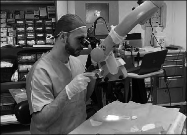
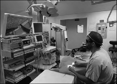
Methods: Participants were recruited among otorhinolaryngology head and neck surgery (OHNS) residents of two tertiary care hospitals. Trainees were asked to complete 4 microvascular end-to-end anastomoses on chicken thighs with the OM and VITOM 3D exoscope. The performances were scored by experienced microvascular surgeons; an objective evaluation of the anastomosis and a subjective assessment of the workload were conducted.
Results: 8 OHNS residents were recruited. Considering the amount of time needed to complete (TTC) the anastomosis, an improvement was shown by all the participants throughout the training program. The objective evaluation of the anastomosis did not show a significant difference. No significant differences were found by analyzing the subjective workload with the different tools.
Conclusions: This article represents the first attempt to compare the use of the OM and the 3D exoscope during training for microsurgery. The results of our study demonstrate the noninferiority of microsurgical training obtained using the 3D exoscope compared to that offered by the OM.

 求助内容:
求助内容: 应助结果提醒方式:


