{"title":"原发性直肠癌伴鳞状细胞癌样分化的异位骨化。","authors":"Yoshihiro Imaeda, Satoshi Arakawa, Hironobu Yasuoka, Hiroyuki Kato, Hidetoshi Nagata, Yukio Asano, Norihiko Kawabe, Kazuya Shiogama, Makoto Urano, Ken-Ichi Inada, Tetsuya Tsukamoto, Akihiko Horiguchi","doi":"10.20407/fmj.2021-013","DOIUrl":null,"url":null,"abstract":"<p><strong>Objectives: </strong>Heterotopic ossification (HO), which occurs when bone tissue forms outside the skeleton, is extremely rare in rectal cancer. Adenocarcinoma is the histological type of all reported primary colorectal cancers with HO. However, in the present case, we observed areas of adenocarcinoma with squamous cell carcinoma-like differentiation. Here we conducted histopathological and immunohistochemical analyses to identify the mechanisms of HO development, to differentiate between adenocarcinoma and squamous cell carcinoma-like phenotypes, and to understand the associated prognostic implications.</p><p><strong>Case report: </strong>A 62-year-old woman was admitted to our hospital with symptoms of intermittent hematochezia without abdominal pain. Colonoscopy revealed stenosis with a protuberant mass in the rectum. Abdominopelvic contrast-enhanced computed tomography showed irregular wall thickness of the rectum, multiple lymph node metastases, and liver metastases. The rectal tumor exhibited calcified deposits with marked hyperintensity. We then performed Hartmann's operation and D3 lymph node resection. The biopsy specimen revealed tubular and solid adenocarcinoma nests and squamous carcinoma-like components over a necrotic extent without secreted mucin. She received chemotherapy (mFOLFOX6 with bevacizumab) as the first option and is alive 5 months after surgery.</p><p><strong>Conclusion: </strong>To the best of our knowledge, this is the first case of heterotopic ossification in a primary rectal cancer with squamous cell carcinoma-like differentiation that was surgically resected. This case suggests that BMP-2 transformed fibroblasts and pluripotent stem cells into osteocytes. We conclude that the squamous cell carcinoma-like lesion was squamous metaplasia of adenocarcinoma.</p>","PeriodicalId":33657,"journal":{"name":"Fujita Medical Journal","volume":"8 4","pages":"134-138"},"PeriodicalIF":0.0000,"publicationDate":"2022-11-01","publicationTypes":"Journal Article","fieldsOfStudy":null,"isOpenAccess":false,"openAccessPdf":"https://www.ncbi.nlm.nih.gov/pmc/articles/PMC9673079/pdf/","citationCount":"0","resultStr":"{\"title\":\"Heterotopic ossification in primary rectal cancer with squamous cell carcinoma-like differentiation.\",\"authors\":\"Yoshihiro Imaeda, Satoshi Arakawa, Hironobu Yasuoka, Hiroyuki Kato, Hidetoshi Nagata, Yukio Asano, Norihiko Kawabe, Kazuya Shiogama, Makoto Urano, Ken-Ichi Inada, Tetsuya Tsukamoto, Akihiko Horiguchi\",\"doi\":\"10.20407/fmj.2021-013\",\"DOIUrl\":null,\"url\":null,\"abstract\":\"<p><strong>Objectives: </strong>Heterotopic ossification (HO), which occurs when bone tissue forms outside the skeleton, is extremely rare in rectal cancer. Adenocarcinoma is the histological type of all reported primary colorectal cancers with HO. However, in the present case, we observed areas of adenocarcinoma with squamous cell carcinoma-like differentiation. Here we conducted histopathological and immunohistochemical analyses to identify the mechanisms of HO development, to differentiate between adenocarcinoma and squamous cell carcinoma-like phenotypes, and to understand the associated prognostic implications.</p><p><strong>Case report: </strong>A 62-year-old woman was admitted to our hospital with symptoms of intermittent hematochezia without abdominal pain. Colonoscopy revealed stenosis with a protuberant mass in the rectum. Abdominopelvic contrast-enhanced computed tomography showed irregular wall thickness of the rectum, multiple lymph node metastases, and liver metastases. The rectal tumor exhibited calcified deposits with marked hyperintensity. We then performed Hartmann's operation and D3 lymph node resection. The biopsy specimen revealed tubular and solid adenocarcinoma nests and squamous carcinoma-like components over a necrotic extent without secreted mucin. She received chemotherapy (mFOLFOX6 with bevacizumab) as the first option and is alive 5 months after surgery.</p><p><strong>Conclusion: </strong>To the best of our knowledge, this is the first case of heterotopic ossification in a primary rectal cancer with squamous cell carcinoma-like differentiation that was surgically resected. This case suggests that BMP-2 transformed fibroblasts and pluripotent stem cells into osteocytes. We conclude that the squamous cell carcinoma-like lesion was squamous metaplasia of adenocarcinoma.</p>\",\"PeriodicalId\":33657,\"journal\":{\"name\":\"Fujita Medical Journal\",\"volume\":\"8 4\",\"pages\":\"134-138\"},\"PeriodicalIF\":0.0000,\"publicationDate\":\"2022-11-01\",\"publicationTypes\":\"Journal Article\",\"fieldsOfStudy\":null,\"isOpenAccess\":false,\"openAccessPdf\":\"https://www.ncbi.nlm.nih.gov/pmc/articles/PMC9673079/pdf/\",\"citationCount\":\"0\",\"resultStr\":null,\"platform\":\"Semanticscholar\",\"paperid\":null,\"PeriodicalName\":\"Fujita Medical Journal\",\"FirstCategoryId\":\"1085\",\"ListUrlMain\":\"https://doi.org/10.20407/fmj.2021-013\",\"RegionNum\":0,\"RegionCategory\":null,\"ArticlePicture\":[],\"TitleCN\":null,\"AbstractTextCN\":null,\"PMCID\":null,\"EPubDate\":\"2022/1/25 0:00:00\",\"PubModel\":\"Epub\",\"JCR\":\"\",\"JCRName\":\"\",\"Score\":null,\"Total\":0}","platform":"Semanticscholar","paperid":null,"PeriodicalName":"Fujita Medical Journal","FirstCategoryId":"1085","ListUrlMain":"https://doi.org/10.20407/fmj.2021-013","RegionNum":0,"RegionCategory":null,"ArticlePicture":[],"TitleCN":null,"AbstractTextCN":null,"PMCID":null,"EPubDate":"2022/1/25 0:00:00","PubModel":"Epub","JCR":"","JCRName":"","Score":null,"Total":0}
Heterotopic ossification in primary rectal cancer with squamous cell carcinoma-like differentiation.
Objectives: Heterotopic ossification (HO), which occurs when bone tissue forms outside the skeleton, is extremely rare in rectal cancer. Adenocarcinoma is the histological type of all reported primary colorectal cancers with HO. However, in the present case, we observed areas of adenocarcinoma with squamous cell carcinoma-like differentiation. Here we conducted histopathological and immunohistochemical analyses to identify the mechanisms of HO development, to differentiate between adenocarcinoma and squamous cell carcinoma-like phenotypes, and to understand the associated prognostic implications.
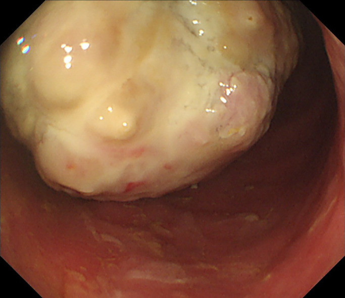
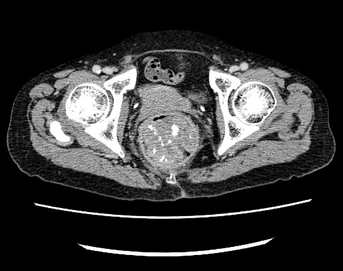
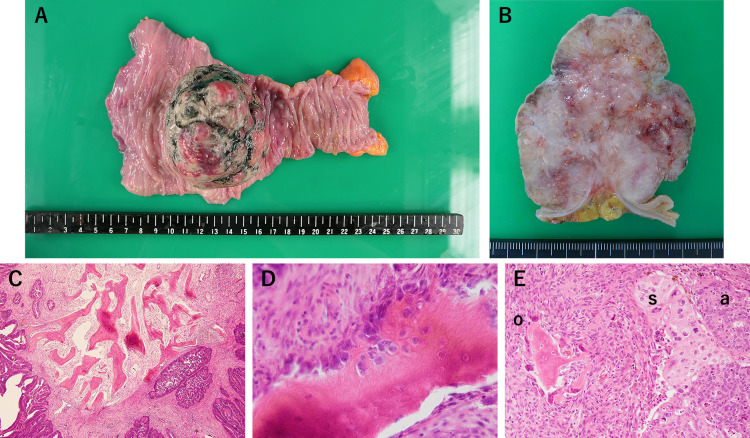
Case report: A 62-year-old woman was admitted to our hospital with symptoms of intermittent hematochezia without abdominal pain. Colonoscopy revealed stenosis with a protuberant mass in the rectum. Abdominopelvic contrast-enhanced computed tomography showed irregular wall thickness of the rectum, multiple lymph node metastases, and liver metastases. The rectal tumor exhibited calcified deposits with marked hyperintensity. We then performed Hartmann's operation and D3 lymph node resection. The biopsy specimen revealed tubular and solid adenocarcinoma nests and squamous carcinoma-like components over a necrotic extent without secreted mucin. She received chemotherapy (mFOLFOX6 with bevacizumab) as the first option and is alive 5 months after surgery.
Conclusion: To the best of our knowledge, this is the first case of heterotopic ossification in a primary rectal cancer with squamous cell carcinoma-like differentiation that was surgically resected. This case suggests that BMP-2 transformed fibroblasts and pluripotent stem cells into osteocytes. We conclude that the squamous cell carcinoma-like lesion was squamous metaplasia of adenocarcinoma.

 求助内容:
求助内容: 应助结果提醒方式:


