Ali Mohamedkhair, Akram Al-Ibraheem, Ahmed Saad Abdlkadir, Omar Jaber
{"title":"乳糜泻合并小肠腺癌患者的FDG PET/CT结果具有挑战性。","authors":"Ali Mohamedkhair, Akram Al-Ibraheem, Ahmed Saad Abdlkadir, Omar Jaber","doi":"10.22038/AOJNMB.2022.61853.1437","DOIUrl":null,"url":null,"abstract":"<p><p>Celiac disease (CD) is a chronic immune-mediated enteropathy that is caused by both environmental (gluten) and genetic (human leukocyte antigen (HLA) and non-HLA genes) factors. Patients may be asymptomatic or exhibit atypical symptoms, necessitating a high index of suspicion for proper diagnosis. The evaluation of CD patients with <sup>18</sup>F-FDG PET/CT imaging can be difficult, owing to the fact that this disease is inflammatory in nature. Typical <sup>18</sup>F-FDG PET/CT gastrointestinal manifestations of celiac disease include increased multifocal or diffuse bowel uptake, whereas single short segmental uptake is rarely encountered; thus, awareness of this wide range of findings is important to guide physicians through proper management and outcome. We report a case of small intestine adenocarcinoma and known CD complaining of recent episodes of diarrhea and weight loss that had a suspicious small bowel wall thickening that corresponds to a short segmental hypermetabolic process on FDG PET/CT follow-up scan. The patient was then referred to the gastroenterology department and underwent a colonoscopy, a biopsy was taken that revealed CD and was negative for malignancy. Furthermore, 6 months later the abovementioned segmental FDG activity was completely resolved without any treatment received at the given time.</p>","PeriodicalId":8503,"journal":{"name":"Asia Oceania Journal of Nuclear Medicine and Biology","volume":"10 2","pages":"155-160"},"PeriodicalIF":0.0000,"publicationDate":"2022-01-01","publicationTypes":"Journal Article","fieldsOfStudy":null,"isOpenAccess":false,"openAccessPdf":"https://www.ncbi.nlm.nih.gov/pmc/articles/PMC9205848/pdf/","citationCount":"0","resultStr":"{\"title\":\"Challenging Results on FDG PET/CT in a Patient with Uncontrolled Celiac Disease and small bowel adenocarcinoma.\",\"authors\":\"Ali Mohamedkhair, Akram Al-Ibraheem, Ahmed Saad Abdlkadir, Omar Jaber\",\"doi\":\"10.22038/AOJNMB.2022.61853.1437\",\"DOIUrl\":null,\"url\":null,\"abstract\":\"<p><p>Celiac disease (CD) is a chronic immune-mediated enteropathy that is caused by both environmental (gluten) and genetic (human leukocyte antigen (HLA) and non-HLA genes) factors. Patients may be asymptomatic or exhibit atypical symptoms, necessitating a high index of suspicion for proper diagnosis. The evaluation of CD patients with <sup>18</sup>F-FDG PET/CT imaging can be difficult, owing to the fact that this disease is inflammatory in nature. Typical <sup>18</sup>F-FDG PET/CT gastrointestinal manifestations of celiac disease include increased multifocal or diffuse bowel uptake, whereas single short segmental uptake is rarely encountered; thus, awareness of this wide range of findings is important to guide physicians through proper management and outcome. We report a case of small intestine adenocarcinoma and known CD complaining of recent episodes of diarrhea and weight loss that had a suspicious small bowel wall thickening that corresponds to a short segmental hypermetabolic process on FDG PET/CT follow-up scan. The patient was then referred to the gastroenterology department and underwent a colonoscopy, a biopsy was taken that revealed CD and was negative for malignancy. Furthermore, 6 months later the abovementioned segmental FDG activity was completely resolved without any treatment received at the given time.</p>\",\"PeriodicalId\":8503,\"journal\":{\"name\":\"Asia Oceania Journal of Nuclear Medicine and Biology\",\"volume\":\"10 2\",\"pages\":\"155-160\"},\"PeriodicalIF\":0.0000,\"publicationDate\":\"2022-01-01\",\"publicationTypes\":\"Journal Article\",\"fieldsOfStudy\":null,\"isOpenAccess\":false,\"openAccessPdf\":\"https://www.ncbi.nlm.nih.gov/pmc/articles/PMC9205848/pdf/\",\"citationCount\":\"0\",\"resultStr\":null,\"platform\":\"Semanticscholar\",\"paperid\":null,\"PeriodicalName\":\"Asia Oceania Journal of Nuclear Medicine and Biology\",\"FirstCategoryId\":\"1085\",\"ListUrlMain\":\"https://doi.org/10.22038/AOJNMB.2022.61853.1437\",\"RegionNum\":0,\"RegionCategory\":null,\"ArticlePicture\":[],\"TitleCN\":null,\"AbstractTextCN\":null,\"PMCID\":null,\"EPubDate\":\"\",\"PubModel\":\"\",\"JCR\":\"Q3\",\"JCRName\":\"Medicine\",\"Score\":null,\"Total\":0}","platform":"Semanticscholar","paperid":null,"PeriodicalName":"Asia Oceania Journal of Nuclear Medicine and Biology","FirstCategoryId":"1085","ListUrlMain":"https://doi.org/10.22038/AOJNMB.2022.61853.1437","RegionNum":0,"RegionCategory":null,"ArticlePicture":[],"TitleCN":null,"AbstractTextCN":null,"PMCID":null,"EPubDate":"","PubModel":"","JCR":"Q3","JCRName":"Medicine","Score":null,"Total":0}
Challenging Results on FDG PET/CT in a Patient with Uncontrolled Celiac Disease and small bowel adenocarcinoma.
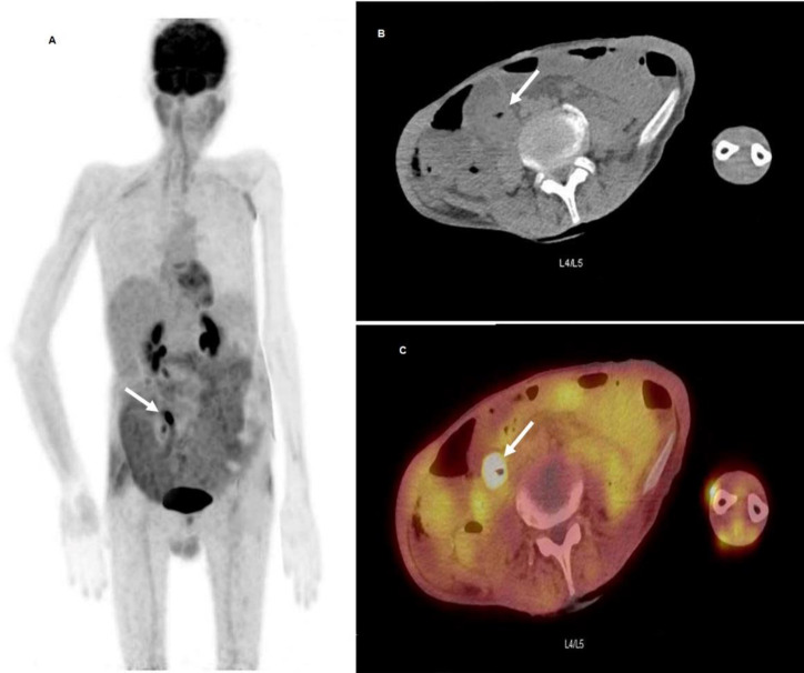
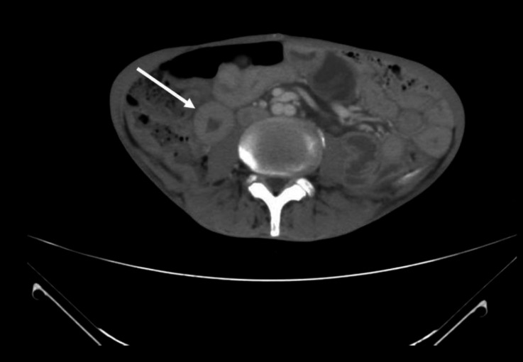
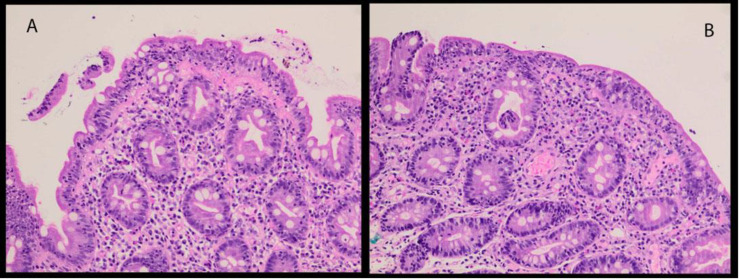
Celiac disease (CD) is a chronic immune-mediated enteropathy that is caused by both environmental (gluten) and genetic (human leukocyte antigen (HLA) and non-HLA genes) factors. Patients may be asymptomatic or exhibit atypical symptoms, necessitating a high index of suspicion for proper diagnosis. The evaluation of CD patients with 18F-FDG PET/CT imaging can be difficult, owing to the fact that this disease is inflammatory in nature. Typical 18F-FDG PET/CT gastrointestinal manifestations of celiac disease include increased multifocal or diffuse bowel uptake, whereas single short segmental uptake is rarely encountered; thus, awareness of this wide range of findings is important to guide physicians through proper management and outcome. We report a case of small intestine adenocarcinoma and known CD complaining of recent episodes of diarrhea and weight loss that had a suspicious small bowel wall thickening that corresponds to a short segmental hypermetabolic process on FDG PET/CT follow-up scan. The patient was then referred to the gastroenterology department and underwent a colonoscopy, a biopsy was taken that revealed CD and was negative for malignancy. Furthermore, 6 months later the abovementioned segmental FDG activity was completely resolved without any treatment received at the given time.

 求助内容:
求助内容: 应助结果提醒方式:


