{"title":"fancd2o通过抑制甾体生成酶和促进小鼠睾丸间质细胞凋亡来减少睾丸激素的产生。","authors":"Xiang Zhai, Xin-Yang Li, Yu-Jing Wang, Ke-Ru Qin, Jin-Rui Hu, Mei-Ning Li, Hai-Long Wang, Rui Guo","doi":"10.3803/EnM.2022.1431","DOIUrl":null,"url":null,"abstract":"<p><strong>Backgruound: </strong>It is well-established that serum testosterone in men decreases with age, yet the underlying mechanism of this change remains elusive.</p><p><strong>Methods: </strong>The expression patterns of Fancd2 opposite-strand (Fancd2os) in BALB/c male mice and testicular tissue derived cell lines (GC-1, GC-2, TM3, and TM4) were assessed using real-time polymerase chain reaction (RT-PCR), Western blot and immunofluorescence. The Fancd2os-overexpressing or knockdown TM3 cells were constructed by infecting them with lentivirus particles and were used to evaluated the function of Fancd2os. The testosterone production was measured using enzyme linked immunosorbent assay (ELISA) and the steroidogenic enzymes such as steroidogenic acute regulatory protein (StAR), P450 cholesterol side-chain cleavage (P450scc), and 3β-hydroxysteroid dehydrogenase (3β-HSD) were analysed using RT-PCR. The apoptosis of TM3 cells induced by ultraviolet light or testicular tissues was detected using flow cytometry, Western blot or dUTP-biotin nick end labeling (TUNEL) assays. Pearson correlation analysis was used to assess the correlation between the Fancd2os expression and TUNEL-positive staining in mouse testicular Leydig cells.</p><p><strong>Results: </strong>The Fancd2os protein was predominantly expressed in mouse testicular Leydig cells and its expression increased with age. Fancd2os overexpression inhibited testosterone levels in TM3 Leydig cells, whereas knockdown of Fancd2os elevated testosterone production. Fancd2os overexpression downregulated the levels of StAR, P450scc and 3β-HSD, while Fancd2os knockdown reversed this effect. Fancd2os overexpression promoted ultraviolet light-induced apoptosis of TM3 cells. In contrast, Fancd2os knockdown restrained apoptosis in TM3 cells. In vivo assays revealed that higher Fancd2os levels and mouse age were associated with increased apoptosis in Leydig cells and decreased serum testosterone levels. Pearson correlation analysis exhibited a strong positive correlation between the expression of Fancd2os and TUNEL-positive staining in mouse testicular Leydig cells.</p><p><strong>Conclusion: </strong>Our findings suggest that Fancd2os regulates testosterone synthesis via both steroidogenic enzymes and the apoptotic pathway.</p>","PeriodicalId":520607,"journal":{"name":"Endocrinology and metabolism (Seoul, Korea)","volume":" ","pages":"533-546"},"PeriodicalIF":4.2000,"publicationDate":"2022-06-01","publicationTypes":"Journal Article","fieldsOfStudy":null,"isOpenAccess":false,"openAccessPdf":"https://ftp.ncbi.nlm.nih.gov/pub/pmc/oa_pdf/13/2a/enm-2022-1431.PMC9262688.pdf","citationCount":"3","resultStr":"{\"title\":\"Fancd2os Reduces Testosterone Production by Inhibiting Steroidogenic Enzymes and Promoting Cellular Apoptosis in Murine Testicular Leydig Cells.\",\"authors\":\"Xiang Zhai, Xin-Yang Li, Yu-Jing Wang, Ke-Ru Qin, Jin-Rui Hu, Mei-Ning Li, Hai-Long Wang, Rui Guo\",\"doi\":\"10.3803/EnM.2022.1431\",\"DOIUrl\":null,\"url\":null,\"abstract\":\"<p><strong>Backgruound: </strong>It is well-established that serum testosterone in men decreases with age, yet the underlying mechanism of this change remains elusive.</p><p><strong>Methods: </strong>The expression patterns of Fancd2 opposite-strand (Fancd2os) in BALB/c male mice and testicular tissue derived cell lines (GC-1, GC-2, TM3, and TM4) were assessed using real-time polymerase chain reaction (RT-PCR), Western blot and immunofluorescence. The Fancd2os-overexpressing or knockdown TM3 cells were constructed by infecting them with lentivirus particles and were used to evaluated the function of Fancd2os. The testosterone production was measured using enzyme linked immunosorbent assay (ELISA) and the steroidogenic enzymes such as steroidogenic acute regulatory protein (StAR), P450 cholesterol side-chain cleavage (P450scc), and 3β-hydroxysteroid dehydrogenase (3β-HSD) were analysed using RT-PCR. The apoptosis of TM3 cells induced by ultraviolet light or testicular tissues was detected using flow cytometry, Western blot or dUTP-biotin nick end labeling (TUNEL) assays. Pearson correlation analysis was used to assess the correlation between the Fancd2os expression and TUNEL-positive staining in mouse testicular Leydig cells.</p><p><strong>Results: </strong>The Fancd2os protein was predominantly expressed in mouse testicular Leydig cells and its expression increased with age. Fancd2os overexpression inhibited testosterone levels in TM3 Leydig cells, whereas knockdown of Fancd2os elevated testosterone production. Fancd2os overexpression downregulated the levels of StAR, P450scc and 3β-HSD, while Fancd2os knockdown reversed this effect. Fancd2os overexpression promoted ultraviolet light-induced apoptosis of TM3 cells. In contrast, Fancd2os knockdown restrained apoptosis in TM3 cells. In vivo assays revealed that higher Fancd2os levels and mouse age were associated with increased apoptosis in Leydig cells and decreased serum testosterone levels. Pearson correlation analysis exhibited a strong positive correlation between the expression of Fancd2os and TUNEL-positive staining in mouse testicular Leydig cells.</p><p><strong>Conclusion: </strong>Our findings suggest that Fancd2os regulates testosterone synthesis via both steroidogenic enzymes and the apoptotic pathway.</p>\",\"PeriodicalId\":520607,\"journal\":{\"name\":\"Endocrinology and metabolism (Seoul, Korea)\",\"volume\":\" \",\"pages\":\"533-546\"},\"PeriodicalIF\":4.2000,\"publicationDate\":\"2022-06-01\",\"publicationTypes\":\"Journal Article\",\"fieldsOfStudy\":null,\"isOpenAccess\":false,\"openAccessPdf\":\"https://ftp.ncbi.nlm.nih.gov/pub/pmc/oa_pdf/13/2a/enm-2022-1431.PMC9262688.pdf\",\"citationCount\":\"3\",\"resultStr\":null,\"platform\":\"Semanticscholar\",\"paperid\":null,\"PeriodicalName\":\"Endocrinology and metabolism (Seoul, Korea)\",\"FirstCategoryId\":\"3\",\"ListUrlMain\":\"https://doi.org/10.3803/EnM.2022.1431\",\"RegionNum\":0,\"RegionCategory\":null,\"ArticlePicture\":[],\"TitleCN\":null,\"AbstractTextCN\":null,\"PMCID\":null,\"EPubDate\":\"2022/6/29 0:00:00\",\"PubModel\":\"Epub\",\"JCR\":\"\",\"JCRName\":\"\",\"Score\":null,\"Total\":0}","platform":"Semanticscholar","paperid":null,"PeriodicalName":"Endocrinology and metabolism (Seoul, Korea)","FirstCategoryId":"3","ListUrlMain":"https://doi.org/10.3803/EnM.2022.1431","RegionNum":0,"RegionCategory":null,"ArticlePicture":[],"TitleCN":null,"AbstractTextCN":null,"PMCID":null,"EPubDate":"2022/6/29 0:00:00","PubModel":"Epub","JCR":"","JCRName":"","Score":null,"Total":0}
Fancd2os Reduces Testosterone Production by Inhibiting Steroidogenic Enzymes and Promoting Cellular Apoptosis in Murine Testicular Leydig Cells.
Backgruound: It is well-established that serum testosterone in men decreases with age, yet the underlying mechanism of this change remains elusive.
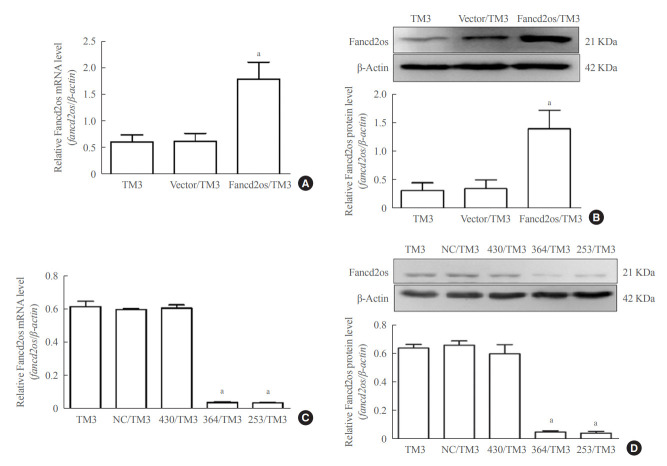
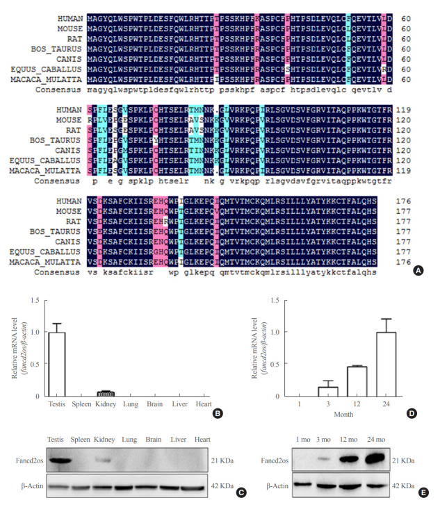
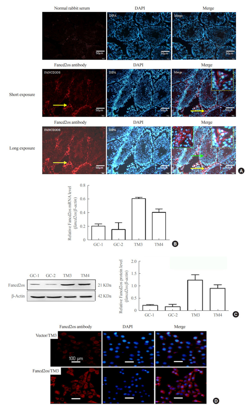
Methods: The expression patterns of Fancd2 opposite-strand (Fancd2os) in BALB/c male mice and testicular tissue derived cell lines (GC-1, GC-2, TM3, and TM4) were assessed using real-time polymerase chain reaction (RT-PCR), Western blot and immunofluorescence. The Fancd2os-overexpressing or knockdown TM3 cells were constructed by infecting them with lentivirus particles and were used to evaluated the function of Fancd2os. The testosterone production was measured using enzyme linked immunosorbent assay (ELISA) and the steroidogenic enzymes such as steroidogenic acute regulatory protein (StAR), P450 cholesterol side-chain cleavage (P450scc), and 3β-hydroxysteroid dehydrogenase (3β-HSD) were analysed using RT-PCR. The apoptosis of TM3 cells induced by ultraviolet light or testicular tissues was detected using flow cytometry, Western blot or dUTP-biotin nick end labeling (TUNEL) assays. Pearson correlation analysis was used to assess the correlation between the Fancd2os expression and TUNEL-positive staining in mouse testicular Leydig cells.
Results: The Fancd2os protein was predominantly expressed in mouse testicular Leydig cells and its expression increased with age. Fancd2os overexpression inhibited testosterone levels in TM3 Leydig cells, whereas knockdown of Fancd2os elevated testosterone production. Fancd2os overexpression downregulated the levels of StAR, P450scc and 3β-HSD, while Fancd2os knockdown reversed this effect. Fancd2os overexpression promoted ultraviolet light-induced apoptosis of TM3 cells. In contrast, Fancd2os knockdown restrained apoptosis in TM3 cells. In vivo assays revealed that higher Fancd2os levels and mouse age were associated with increased apoptosis in Leydig cells and decreased serum testosterone levels. Pearson correlation analysis exhibited a strong positive correlation between the expression of Fancd2os and TUNEL-positive staining in mouse testicular Leydig cells.
Conclusion: Our findings suggest that Fancd2os regulates testosterone synthesis via both steroidogenic enzymes and the apoptotic pathway.

 求助内容:
求助内容: 应助结果提醒方式:


