Raffaele Aspide, Giacomo Bertolini, Laura Maria Beatrice Belotti, Luca Albini Riccioli, Francesco Toni, Diego Mazzatenta, Giorgio Palandri, Luigi Vetrugno, Daniele Guerino Biasucci
{"title":"使用彩色多普勒评估视神经鞘直径的CLOSED方案:特发性常压脑积水患者队列的比较研究。","authors":"Raffaele Aspide, Giacomo Bertolini, Laura Maria Beatrice Belotti, Luca Albini Riccioli, Francesco Toni, Diego Mazzatenta, Giorgio Palandri, Luigi Vetrugno, Daniele Guerino Biasucci","doi":"10.1186/s13089-022-00291-5","DOIUrl":null,"url":null,"abstract":"<p><strong>Background: </strong>Sonographic assessment of the optic nerve sheath diameter represents a promising non-invasive technique for estimation of the intracranial pressure. A wide inter-observer variability, along with a lack of a standardized protocol for the optic nerve sheath diameter measurements, could lead to over- or under-estimation. The present study was aimed at evaluating feasibility of color-Doppler for better delineating optic nerve sheath borders, comparing it to B-mode imaging, using the magnetic resonance measurements as a comparison.</p><p><strong>Methods: </strong>Optic nerve sheath diameters were evaluated using magnetic resonance by an expert radiologist in a cohort of patients with suspected idiopathic normal pressure hydrocephalus. Magnetic resonance findings were evaluated twice. In the first half of this cohort, optic nerve sheath diameters were measured using B-mode only, in the second half applying color-Doppler. Measurements obtained using these two techniques were compared to magnetic resonance imaging measurements. The Bland-Altman analysis and concordance correlation coefficient were computed to quantify the strength of agreement between the two magnetic resonance assessments. Box plots and average (± SD) were used to compare assessments by sonographic and magnetic resonance methods.</p><p><strong>Results: </strong>Fifty patients were included. MRI assessment showed a moderate concordance correlation coefficient. Optic nerve sheath diameters measured applying color-Doppler were lower (p < 0.001) and less scattered compared to B-mode assessment, which approached more to magnetic resonance measurements.</p><p><strong>Conclusions: </strong>In this cohort of patients, magnetic resonance showed high intra-rater variability in optic nerve sheath diameter assessments. Optic nerve sheath diameter assessments using color-Doppler yielded lower and less scattered diameters compared to B-mode only.</p>","PeriodicalId":36911,"journal":{"name":"Ultrasound Journal","volume":" ","pages":"43"},"PeriodicalIF":2.9000,"publicationDate":"2022-10-29","publicationTypes":"Journal Article","fieldsOfStudy":null,"isOpenAccess":false,"openAccessPdf":"https://www.ncbi.nlm.nih.gov/pmc/articles/PMC9617984/pdf/","citationCount":"9","resultStr":"{\"title\":\"The CLOSED protocol to assess optic nerve sheath diameter using color-Doppler: a comparison study in a cohort of idiopathic normal pressure hydrocephalus patients.\",\"authors\":\"Raffaele Aspide, Giacomo Bertolini, Laura Maria Beatrice Belotti, Luca Albini Riccioli, Francesco Toni, Diego Mazzatenta, Giorgio Palandri, Luigi Vetrugno, Daniele Guerino Biasucci\",\"doi\":\"10.1186/s13089-022-00291-5\",\"DOIUrl\":null,\"url\":null,\"abstract\":\"<p><strong>Background: </strong>Sonographic assessment of the optic nerve sheath diameter represents a promising non-invasive technique for estimation of the intracranial pressure. A wide inter-observer variability, along with a lack of a standardized protocol for the optic nerve sheath diameter measurements, could lead to over- or under-estimation. The present study was aimed at evaluating feasibility of color-Doppler for better delineating optic nerve sheath borders, comparing it to B-mode imaging, using the magnetic resonance measurements as a comparison.</p><p><strong>Methods: </strong>Optic nerve sheath diameters were evaluated using magnetic resonance by an expert radiologist in a cohort of patients with suspected idiopathic normal pressure hydrocephalus. Magnetic resonance findings were evaluated twice. In the first half of this cohort, optic nerve sheath diameters were measured using B-mode only, in the second half applying color-Doppler. Measurements obtained using these two techniques were compared to magnetic resonance imaging measurements. The Bland-Altman analysis and concordance correlation coefficient were computed to quantify the strength of agreement between the two magnetic resonance assessments. Box plots and average (± SD) were used to compare assessments by sonographic and magnetic resonance methods.</p><p><strong>Results: </strong>Fifty patients were included. MRI assessment showed a moderate concordance correlation coefficient. Optic nerve sheath diameters measured applying color-Doppler were lower (p < 0.001) and less scattered compared to B-mode assessment, which approached more to magnetic resonance measurements.</p><p><strong>Conclusions: </strong>In this cohort of patients, magnetic resonance showed high intra-rater variability in optic nerve sheath diameter assessments. Optic nerve sheath diameter assessments using color-Doppler yielded lower and less scattered diameters compared to B-mode only.</p>\",\"PeriodicalId\":36911,\"journal\":{\"name\":\"Ultrasound Journal\",\"volume\":\" \",\"pages\":\"43\"},\"PeriodicalIF\":2.9000,\"publicationDate\":\"2022-10-29\",\"publicationTypes\":\"Journal Article\",\"fieldsOfStudy\":null,\"isOpenAccess\":false,\"openAccessPdf\":\"https://www.ncbi.nlm.nih.gov/pmc/articles/PMC9617984/pdf/\",\"citationCount\":\"9\",\"resultStr\":null,\"platform\":\"Semanticscholar\",\"paperid\":null,\"PeriodicalName\":\"Ultrasound Journal\",\"FirstCategoryId\":\"1085\",\"ListUrlMain\":\"https://doi.org/10.1186/s13089-022-00291-5\",\"RegionNum\":0,\"RegionCategory\":null,\"ArticlePicture\":[],\"TitleCN\":null,\"AbstractTextCN\":null,\"PMCID\":null,\"EPubDate\":\"\",\"PubModel\":\"\",\"JCR\":\"Q2\",\"JCRName\":\"Medicine\",\"Score\":null,\"Total\":0}","platform":"Semanticscholar","paperid":null,"PeriodicalName":"Ultrasound Journal","FirstCategoryId":"1085","ListUrlMain":"https://doi.org/10.1186/s13089-022-00291-5","RegionNum":0,"RegionCategory":null,"ArticlePicture":[],"TitleCN":null,"AbstractTextCN":null,"PMCID":null,"EPubDate":"","PubModel":"","JCR":"Q2","JCRName":"Medicine","Score":null,"Total":0}
The CLOSED protocol to assess optic nerve sheath diameter using color-Doppler: a comparison study in a cohort of idiopathic normal pressure hydrocephalus patients.
Background: Sonographic assessment of the optic nerve sheath diameter represents a promising non-invasive technique for estimation of the intracranial pressure. A wide inter-observer variability, along with a lack of a standardized protocol for the optic nerve sheath diameter measurements, could lead to over- or under-estimation. The present study was aimed at evaluating feasibility of color-Doppler for better delineating optic nerve sheath borders, comparing it to B-mode imaging, using the magnetic resonance measurements as a comparison.
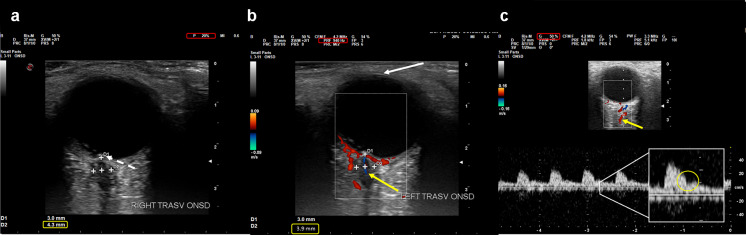
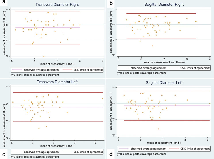
Methods: Optic nerve sheath diameters were evaluated using magnetic resonance by an expert radiologist in a cohort of patients with suspected idiopathic normal pressure hydrocephalus. Magnetic resonance findings were evaluated twice. In the first half of this cohort, optic nerve sheath diameters were measured using B-mode only, in the second half applying color-Doppler. Measurements obtained using these two techniques were compared to magnetic resonance imaging measurements. The Bland-Altman analysis and concordance correlation coefficient were computed to quantify the strength of agreement between the two magnetic resonance assessments. Box plots and average (± SD) were used to compare assessments by sonographic and magnetic resonance methods.
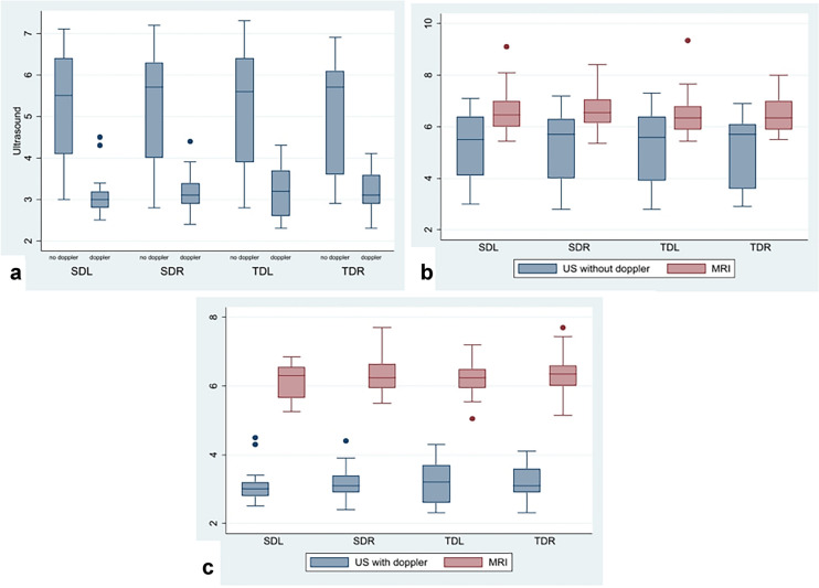
Results: Fifty patients were included. MRI assessment showed a moderate concordance correlation coefficient. Optic nerve sheath diameters measured applying color-Doppler were lower (p < 0.001) and less scattered compared to B-mode assessment, which approached more to magnetic resonance measurements.
Conclusions: In this cohort of patients, magnetic resonance showed high intra-rater variability in optic nerve sheath diameter assessments. Optic nerve sheath diameter assessments using color-Doppler yielded lower and less scattered diameters compared to B-mode only.

 求助内容:
求助内容: 应助结果提醒方式:


