{"title":"膝关节霍金斯征:一种不寻常的废用性骨质减少的影像学表现和临床意义。","authors":"Nicholas C Adams, Robin L Alonge, Lance D Edmonds","doi":"10.25259/JCIS_33_2022","DOIUrl":null,"url":null,"abstract":"<p><p>Disuse osteopenia (DO) is a disorder due to reduced weight-bearing often following immobilization injuries. It is most commonly observed in the ankles and knees and is believed to be due primarily to increased bone reabsorption associated with disuse. Both traditional radiography and magnetic resonance (MR) imaging are useful in identifying abnormalities associated with DO. Specifically, linear subchondral osteopenia has been given the term \"Hawkins sign\" when seen in the talus, but this finding may also be seen elsewhere. When present, it not only is an indication of DO but also indicates the presence of sufficient vascular flow, and the unlikely development of avascular necrosis. We report a case of Hawkins sign of the knee demonstrated on radiography and MR and demonstrate the clinical importance of recognizing this sign, outside its usual setting, in assessing the prognosis of a healing fracture.</p>","PeriodicalId":15512,"journal":{"name":"Journal of Clinical Imaging Science","volume":" ","pages":"51"},"PeriodicalIF":1.1000,"publicationDate":"2022-08-22","publicationTypes":"Journal Article","fieldsOfStudy":null,"isOpenAccess":false,"openAccessPdf":"https://ftp.ncbi.nlm.nih.gov/pub/pmc/oa_pdf/d9/4d/JCIS-12-51.PMC9479503.pdf","citationCount":"0","resultStr":"{\"title\":\"Hawkins sign of the knee: Imaging appearance and clinical implication of an unusual pattern of disuse osteopenia.\",\"authors\":\"Nicholas C Adams, Robin L Alonge, Lance D Edmonds\",\"doi\":\"10.25259/JCIS_33_2022\",\"DOIUrl\":null,\"url\":null,\"abstract\":\"<p><p>Disuse osteopenia (DO) is a disorder due to reduced weight-bearing often following immobilization injuries. It is most commonly observed in the ankles and knees and is believed to be due primarily to increased bone reabsorption associated with disuse. Both traditional radiography and magnetic resonance (MR) imaging are useful in identifying abnormalities associated with DO. Specifically, linear subchondral osteopenia has been given the term \\\"Hawkins sign\\\" when seen in the talus, but this finding may also be seen elsewhere. When present, it not only is an indication of DO but also indicates the presence of sufficient vascular flow, and the unlikely development of avascular necrosis. We report a case of Hawkins sign of the knee demonstrated on radiography and MR and demonstrate the clinical importance of recognizing this sign, outside its usual setting, in assessing the prognosis of a healing fracture.</p>\",\"PeriodicalId\":15512,\"journal\":{\"name\":\"Journal of Clinical Imaging Science\",\"volume\":\" \",\"pages\":\"51\"},\"PeriodicalIF\":1.1000,\"publicationDate\":\"2022-08-22\",\"publicationTypes\":\"Journal Article\",\"fieldsOfStudy\":null,\"isOpenAccess\":false,\"openAccessPdf\":\"https://ftp.ncbi.nlm.nih.gov/pub/pmc/oa_pdf/d9/4d/JCIS-12-51.PMC9479503.pdf\",\"citationCount\":\"0\",\"resultStr\":null,\"platform\":\"Semanticscholar\",\"paperid\":null,\"PeriodicalName\":\"Journal of Clinical Imaging Science\",\"FirstCategoryId\":\"1085\",\"ListUrlMain\":\"https://doi.org/10.25259/JCIS_33_2022\",\"RegionNum\":0,\"RegionCategory\":null,\"ArticlePicture\":[],\"TitleCN\":null,\"AbstractTextCN\":null,\"PMCID\":null,\"EPubDate\":\"2022/1/1 0:00:00\",\"PubModel\":\"eCollection\",\"JCR\":\"Q3\",\"JCRName\":\"RADIOLOGY, NUCLEAR MEDICINE & MEDICAL IMAGING\",\"Score\":null,\"Total\":0}","platform":"Semanticscholar","paperid":null,"PeriodicalName":"Journal of Clinical Imaging Science","FirstCategoryId":"1085","ListUrlMain":"https://doi.org/10.25259/JCIS_33_2022","RegionNum":0,"RegionCategory":null,"ArticlePicture":[],"TitleCN":null,"AbstractTextCN":null,"PMCID":null,"EPubDate":"2022/1/1 0:00:00","PubModel":"eCollection","JCR":"Q3","JCRName":"RADIOLOGY, NUCLEAR MEDICINE & MEDICAL IMAGING","Score":null,"Total":0}
Hawkins sign of the knee: Imaging appearance and clinical implication of an unusual pattern of disuse osteopenia.
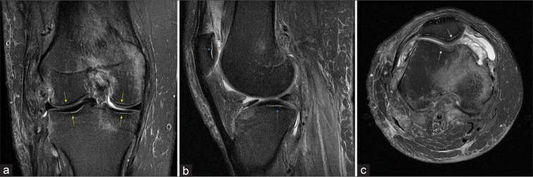
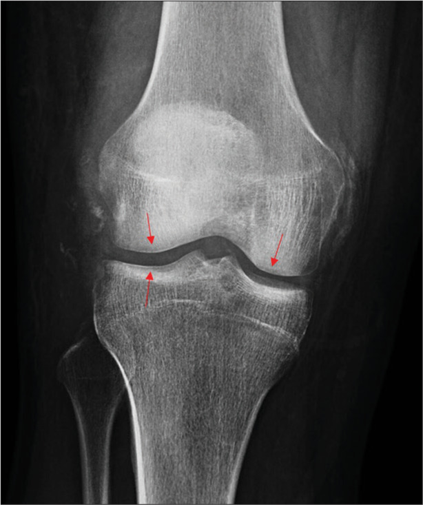
Disuse osteopenia (DO) is a disorder due to reduced weight-bearing often following immobilization injuries. It is most commonly observed in the ankles and knees and is believed to be due primarily to increased bone reabsorption associated with disuse. Both traditional radiography and magnetic resonance (MR) imaging are useful in identifying abnormalities associated with DO. Specifically, linear subchondral osteopenia has been given the term "Hawkins sign" when seen in the talus, but this finding may also be seen elsewhere. When present, it not only is an indication of DO but also indicates the presence of sufficient vascular flow, and the unlikely development of avascular necrosis. We report a case of Hawkins sign of the knee demonstrated on radiography and MR and demonstrate the clinical importance of recognizing this sign, outside its usual setting, in assessing the prognosis of a healing fracture.
期刊介绍:
The Journal of Clinical Imaging Science (JCIS) is an open access peer-reviewed journal committed to publishing high-quality articles in the field of Imaging Science. The journal aims to present Imaging Science and relevant clinical information in an understandable and useful format. The journal is owned and published by the Scientific Scholar. Audience Our audience includes Radiologists, Researchers, Clinicians, medical professionals and students. Review process JCIS has a highly rigorous peer-review process that makes sure that manuscripts are scientifically accurate, relevant, novel and important. Authors disclose all conflicts, affiliations and financial associations such that the published content is not biased.

 求助内容:
求助内容: 应助结果提醒方式:


