{"title":"骨通道窗口大小和窗口上的胶原屏障对窦底抬高的影响:兔窦模型的临床前研究。","authors":"Jeong-Eun Sim, Sangyup Kim, Ji-Youn Hong, Seung-Il Shin, Jong-Hyuk Chung, Hyun-Chang Lim","doi":"10.5051/jpis.2105560278","DOIUrl":null,"url":null,"abstract":"<p><strong>Purpose: </strong>The aim of this study was to investigate the effect of (1) the size of the bony access window and (2) collagen membrane coverage over the window in sinus floor elevation in a rabbit sinus model.</p><p><strong>Methods: </strong>Small bony access windows (SW; ø 2.8 mm) were made in 6 rabbits and large windows (LW; ø 6 mm) in 6 other rabbits. Both sinuses in each rabbit were allocated to groups with or without coverage of a collagen membrane (CM) on the window, resulting in 4 groups: SW, LW, SW+CM, and LW+CM. After 4 weeks of healing, micro-computed tomographic, histologic, and histomorphometric analyses were performed.</p><p><strong>Results: </strong>Bony healing in the window area was incomplete in all groups, but most bone graft particles were well confined in the augmented cavity. Histologically, the pattern of new bone formation was similar in all groups. Histomorphometrically, the percentage of newly formed bone was greater in the groups with CM than in the groups without CM, and in the groups with SW than in the groups with LW (12.92%±6.40% in the SW+CM group, 4.21%±7.73% in the SW group, 10.45%±4.81% in the LW+CM group, 11.77%±3.83% in the LW group). The above differences were not statistically significant (<i>P></i>0.05).</p><p><strong>Conclusions: </strong>The combination of a small bony access window and the use of a collagen membrane over the window favored new bone formation compared to other groups, but this result should be further investigated due to the limitations of the present animal model.</p>","PeriodicalId":520685,"journal":{"name":"Journal of periodontal & implant science","volume":" ","pages":"325-337"},"PeriodicalIF":3.2000,"publicationDate":"2022-08-01","publicationTypes":"Journal Article","fieldsOfStudy":null,"isOpenAccess":false,"openAccessPdf":"https://ftp.ncbi.nlm.nih.gov/pub/pmc/oa_pdf/b5/ea/jpis-52-325.PMC9436639.pdf","citationCount":"3","resultStr":"{\"title\":\"Effect of the size of the bony access window and the collagen barrier over the window in sinus floor elevation: a preclinical investigation in a rabbit sinus model.\",\"authors\":\"Jeong-Eun Sim, Sangyup Kim, Ji-Youn Hong, Seung-Il Shin, Jong-Hyuk Chung, Hyun-Chang Lim\",\"doi\":\"10.5051/jpis.2105560278\",\"DOIUrl\":null,\"url\":null,\"abstract\":\"<p><strong>Purpose: </strong>The aim of this study was to investigate the effect of (1) the size of the bony access window and (2) collagen membrane coverage over the window in sinus floor elevation in a rabbit sinus model.</p><p><strong>Methods: </strong>Small bony access windows (SW; ø 2.8 mm) were made in 6 rabbits and large windows (LW; ø 6 mm) in 6 other rabbits. Both sinuses in each rabbit were allocated to groups with or without coverage of a collagen membrane (CM) on the window, resulting in 4 groups: SW, LW, SW+CM, and LW+CM. After 4 weeks of healing, micro-computed tomographic, histologic, and histomorphometric analyses were performed.</p><p><strong>Results: </strong>Bony healing in the window area was incomplete in all groups, but most bone graft particles were well confined in the augmented cavity. Histologically, the pattern of new bone formation was similar in all groups. Histomorphometrically, the percentage of newly formed bone was greater in the groups with CM than in the groups without CM, and in the groups with SW than in the groups with LW (12.92%±6.40% in the SW+CM group, 4.21%±7.73% in the SW group, 10.45%±4.81% in the LW+CM group, 11.77%±3.83% in the LW group). The above differences were not statistically significant (<i>P></i>0.05).</p><p><strong>Conclusions: </strong>The combination of a small bony access window and the use of a collagen membrane over the window favored new bone formation compared to other groups, but this result should be further investigated due to the limitations of the present animal model.</p>\",\"PeriodicalId\":520685,\"journal\":{\"name\":\"Journal of periodontal & implant science\",\"volume\":\" \",\"pages\":\"325-337\"},\"PeriodicalIF\":3.2000,\"publicationDate\":\"2022-08-01\",\"publicationTypes\":\"Journal Article\",\"fieldsOfStudy\":null,\"isOpenAccess\":false,\"openAccessPdf\":\"https://ftp.ncbi.nlm.nih.gov/pub/pmc/oa_pdf/b5/ea/jpis-52-325.PMC9436639.pdf\",\"citationCount\":\"3\",\"resultStr\":null,\"platform\":\"Semanticscholar\",\"paperid\":null,\"PeriodicalName\":\"Journal of periodontal & implant science\",\"FirstCategoryId\":\"3\",\"ListUrlMain\":\"https://doi.org/10.5051/jpis.2105560278\",\"RegionNum\":0,\"RegionCategory\":null,\"ArticlePicture\":[],\"TitleCN\":null,\"AbstractTextCN\":null,\"PMCID\":null,\"EPubDate\":\"\",\"PubModel\":\"\",\"JCR\":\"\",\"JCRName\":\"\",\"Score\":null,\"Total\":0}","platform":"Semanticscholar","paperid":null,"PeriodicalName":"Journal of periodontal & implant science","FirstCategoryId":"3","ListUrlMain":"https://doi.org/10.5051/jpis.2105560278","RegionNum":0,"RegionCategory":null,"ArticlePicture":[],"TitleCN":null,"AbstractTextCN":null,"PMCID":null,"EPubDate":"","PubModel":"","JCR":"","JCRName":"","Score":null,"Total":0}
Effect of the size of the bony access window and the collagen barrier over the window in sinus floor elevation: a preclinical investigation in a rabbit sinus model.
Purpose: The aim of this study was to investigate the effect of (1) the size of the bony access window and (2) collagen membrane coverage over the window in sinus floor elevation in a rabbit sinus model.
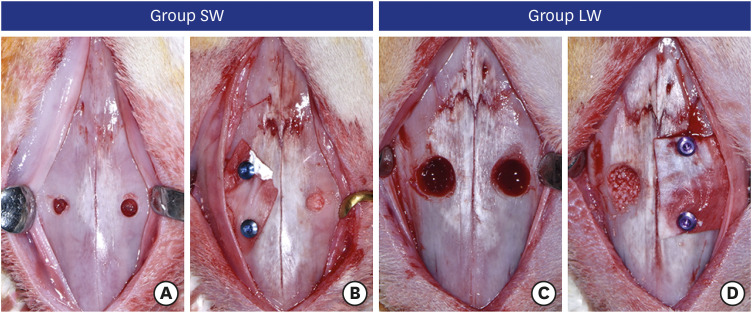
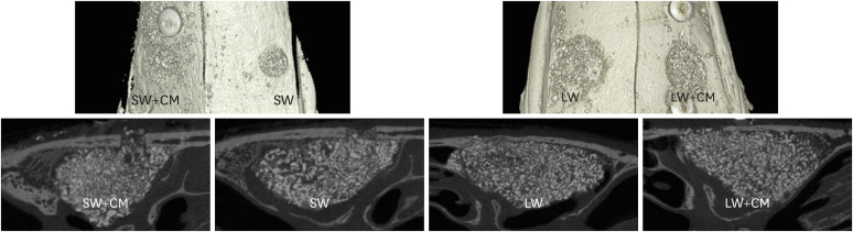
Methods: Small bony access windows (SW; ø 2.8 mm) were made in 6 rabbits and large windows (LW; ø 6 mm) in 6 other rabbits. Both sinuses in each rabbit were allocated to groups with or without coverage of a collagen membrane (CM) on the window, resulting in 4 groups: SW, LW, SW+CM, and LW+CM. After 4 weeks of healing, micro-computed tomographic, histologic, and histomorphometric analyses were performed.
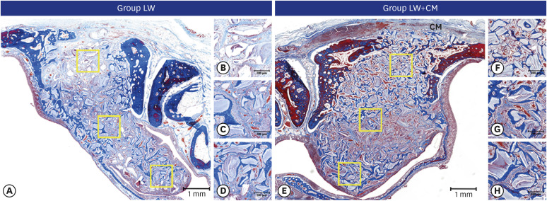
Results: Bony healing in the window area was incomplete in all groups, but most bone graft particles were well confined in the augmented cavity. Histologically, the pattern of new bone formation was similar in all groups. Histomorphometrically, the percentage of newly formed bone was greater in the groups with CM than in the groups without CM, and in the groups with SW than in the groups with LW (12.92%±6.40% in the SW+CM group, 4.21%±7.73% in the SW group, 10.45%±4.81% in the LW+CM group, 11.77%±3.83% in the LW group). The above differences were not statistically significant (P>0.05).
Conclusions: The combination of a small bony access window and the use of a collagen membrane over the window favored new bone formation compared to other groups, but this result should be further investigated due to the limitations of the present animal model.

 求助内容:
求助内容: 应助结果提醒方式:


