{"title":"免疫荧光信号强度测量作为半定量工具来评估肌肉活检中肌聚糖复合物的表达。","authors":"Simona Zanotti, Francesca Magri, Francesca Poggetti, Michela Ripolone, Daniele Velardo, Francesco Fortunato, Patrizia Ciscato, Maurizio Moggio, Stefania Corti, Giacomo Pietro Comi, Monica Sciacco","doi":"10.4081/ejh.2022.3418","DOIUrl":null,"url":null,"abstract":"<p><p>Sarcoglycanopathies are highly heterogeneous in terms of disease progression, muscular weakness, loss of ambulation and cardiac/respiratory involvement. Their clinical severity usually correlates with the residual protein amount, which makes protein quantification extremely relevant. Sarcoglycanopathy diagnosis is genetic, but skeletal muscle analysis - by both immunohistochemistry and Western blot (WB) - is still mandatory to establish the correct diagnostic process. Unfortunately, however, WB analysis cannot be performed if the bioptic specimen is scarce. This study provides a sensitive tool for semi-quantification of residual amount of sarcoglycans in patients affected by sarcoglycanopathies, based on immunofluorescence staining on skeletal muscle sections, image acquisition and software elaboration. We applied this method to eleven sarcoglycanopathies, seven Becker muscular dystrophies and four age-matched controls. Fluorescence data analysed in patients and compared to age-matched controls showed a significant reduction of the mutated sarcoglycan expression and a variable reduction of the other sarcoglycans. Fluorescence normalized data analysed in relation to the age of onset of the disease, showed a negative correlation of α-sarcoglycan fluorescent signal versus fibrosis in patients with an early age of onset and a negative correlation between δ-sarcoglycan signal and fibrosis in both intermediate and late age of onset groups. The availability of a method that allows objective quantification of the sarcolemmal proteins, faster and less consuming than WB analysis and able to detect low residual sarcoglycan expression with great sensitivity, proves useful to better define both patient prognosis and expected disease evolution. The proposed method could be employed also to monitor the efficacy of therapeutic interventions and during clinical trials.</p>","PeriodicalId":50487,"journal":{"name":"European Journal of Histochemistry","volume":null,"pages":null},"PeriodicalIF":2.1000,"publicationDate":"2022-09-01","publicationTypes":"Journal Article","fieldsOfStudy":null,"isOpenAccess":false,"openAccessPdf":"https://ftp.ncbi.nlm.nih.gov/pub/pmc/oa_pdf/ca/42/ejh-66-3-3418.PMC9471914.pdf","citationCount":"0","resultStr":"{\"title\":\"Immunofluorescence signal intensity measurements as a semi-quantitative tool to assess sarcoglycan complex expression in muscle biopsy.\",\"authors\":\"Simona Zanotti, Francesca Magri, Francesca Poggetti, Michela Ripolone, Daniele Velardo, Francesco Fortunato, Patrizia Ciscato, Maurizio Moggio, Stefania Corti, Giacomo Pietro Comi, Monica Sciacco\",\"doi\":\"10.4081/ejh.2022.3418\",\"DOIUrl\":null,\"url\":null,\"abstract\":\"<p><p>Sarcoglycanopathies are highly heterogeneous in terms of disease progression, muscular weakness, loss of ambulation and cardiac/respiratory involvement. Their clinical severity usually correlates with the residual protein amount, which makes protein quantification extremely relevant. Sarcoglycanopathy diagnosis is genetic, but skeletal muscle analysis - by both immunohistochemistry and Western blot (WB) - is still mandatory to establish the correct diagnostic process. Unfortunately, however, WB analysis cannot be performed if the bioptic specimen is scarce. This study provides a sensitive tool for semi-quantification of residual amount of sarcoglycans in patients affected by sarcoglycanopathies, based on immunofluorescence staining on skeletal muscle sections, image acquisition and software elaboration. We applied this method to eleven sarcoglycanopathies, seven Becker muscular dystrophies and four age-matched controls. Fluorescence data analysed in patients and compared to age-matched controls showed a significant reduction of the mutated sarcoglycan expression and a variable reduction of the other sarcoglycans. Fluorescence normalized data analysed in relation to the age of onset of the disease, showed a negative correlation of α-sarcoglycan fluorescent signal versus fibrosis in patients with an early age of onset and a negative correlation between δ-sarcoglycan signal and fibrosis in both intermediate and late age of onset groups. The availability of a method that allows objective quantification of the sarcolemmal proteins, faster and less consuming than WB analysis and able to detect low residual sarcoglycan expression with great sensitivity, proves useful to better define both patient prognosis and expected disease evolution. The proposed method could be employed also to monitor the efficacy of therapeutic interventions and during clinical trials.</p>\",\"PeriodicalId\":50487,\"journal\":{\"name\":\"European Journal of Histochemistry\",\"volume\":null,\"pages\":null},\"PeriodicalIF\":2.1000,\"publicationDate\":\"2022-09-01\",\"publicationTypes\":\"Journal Article\",\"fieldsOfStudy\":null,\"isOpenAccess\":false,\"openAccessPdf\":\"https://ftp.ncbi.nlm.nih.gov/pub/pmc/oa_pdf/ca/42/ejh-66-3-3418.PMC9471914.pdf\",\"citationCount\":\"0\",\"resultStr\":null,\"platform\":\"Semanticscholar\",\"paperid\":null,\"PeriodicalName\":\"European Journal of Histochemistry\",\"FirstCategoryId\":\"99\",\"ListUrlMain\":\"https://doi.org/10.4081/ejh.2022.3418\",\"RegionNum\":4,\"RegionCategory\":\"生物学\",\"ArticlePicture\":[],\"TitleCN\":null,\"AbstractTextCN\":null,\"PMCID\":null,\"EPubDate\":\"\",\"PubModel\":\"\",\"JCR\":\"Q4\",\"JCRName\":\"CELL BIOLOGY\",\"Score\":null,\"Total\":0}","platform":"Semanticscholar","paperid":null,"PeriodicalName":"European Journal of Histochemistry","FirstCategoryId":"99","ListUrlMain":"https://doi.org/10.4081/ejh.2022.3418","RegionNum":4,"RegionCategory":"生物学","ArticlePicture":[],"TitleCN":null,"AbstractTextCN":null,"PMCID":null,"EPubDate":"","PubModel":"","JCR":"Q4","JCRName":"CELL BIOLOGY","Score":null,"Total":0}
Immunofluorescence signal intensity measurements as a semi-quantitative tool to assess sarcoglycan complex expression in muscle biopsy.
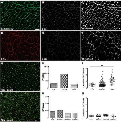
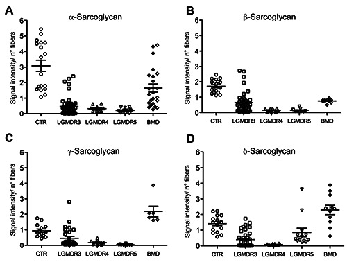
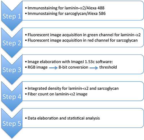
Sarcoglycanopathies are highly heterogeneous in terms of disease progression, muscular weakness, loss of ambulation and cardiac/respiratory involvement. Their clinical severity usually correlates with the residual protein amount, which makes protein quantification extremely relevant. Sarcoglycanopathy diagnosis is genetic, but skeletal muscle analysis - by both immunohistochemistry and Western blot (WB) - is still mandatory to establish the correct diagnostic process. Unfortunately, however, WB analysis cannot be performed if the bioptic specimen is scarce. This study provides a sensitive tool for semi-quantification of residual amount of sarcoglycans in patients affected by sarcoglycanopathies, based on immunofluorescence staining on skeletal muscle sections, image acquisition and software elaboration. We applied this method to eleven sarcoglycanopathies, seven Becker muscular dystrophies and four age-matched controls. Fluorescence data analysed in patients and compared to age-matched controls showed a significant reduction of the mutated sarcoglycan expression and a variable reduction of the other sarcoglycans. Fluorescence normalized data analysed in relation to the age of onset of the disease, showed a negative correlation of α-sarcoglycan fluorescent signal versus fibrosis in patients with an early age of onset and a negative correlation between δ-sarcoglycan signal and fibrosis in both intermediate and late age of onset groups. The availability of a method that allows objective quantification of the sarcolemmal proteins, faster and less consuming than WB analysis and able to detect low residual sarcoglycan expression with great sensitivity, proves useful to better define both patient prognosis and expected disease evolution. The proposed method could be employed also to monitor the efficacy of therapeutic interventions and during clinical trials.
期刊介绍:
The Journal publishes original papers concerning investigations by histochemical and immunohistochemical methods, and performed with the aid of light, super-resolution and electron microscopy, cytometry and imaging techniques. Coverage extends to:
functional cell and tissue biology in animals and plants;
cell differentiation and death;
cell-cell interaction and molecular trafficking;
biology of cell development and senescence;
nerve and muscle cell biology;
cellular basis of diseases.
The histochemical approach is nowadays essentially aimed at locating molecules in the very place where they exert their biological roles, and at describing dynamically specific chemical activities in living cells. Basic research on cell functional organization is essential for understanding the mechanisms underlying major biological processes such as differentiation, the control of tissue homeostasis, and the regulation of normal and tumor cell growth. Even more than in the past, the European Journal of Histochemistry, as a journal of functional cytology, represents the venue where cell scientists may present and discuss their original results, technical improvements and theories.
 求助内容:
求助内容: 应助结果提醒方式:


