Alimu Keremu, Pazila Aila, Aikebaier Tusun, Maimaitiaili Abulikemu, Xiaoguang Zou
{"title":"骨间充质干细胞的细胞外囊泡将microRNA-206转运到骨肉瘤细胞中,并靶向NRSN2阻断ERK1/2-Bcl-xL信号通路。","authors":"Alimu Keremu, Pazila Aila, Aikebaier Tusun, Maimaitiaili Abulikemu, Xiaoguang Zou","doi":"10.4081/ejh.2022.3394","DOIUrl":null,"url":null,"abstract":"<p><p>Osteosarcoma (OS) is a kind of malignant tumor originating from mesenchymal tissue Bone mesenchymal stem cells-derived extracellular vesicles (BMSCs-EVs) can play important roles in OS. This study investigated the mechanism of BMSCs-EVs on OS. BMSC surface antigens and adipogenic and osteogenic differentiation were detected by flow cytometry, and oil red O and alizarin red staining. EVs were isolated from BMSCs by differential centrifugation and identified by transmission electron microscopy, nanoparticle tracking analysis, and Western blot (WB). miR-206 and neurensin-2 (NRSN2) levels in human osteoblast hFOB 1.19 or OS cells (143B, MG-63, Saos2, HOS) were detected by RT-qPCR. Human OS cells with lower miR-206 levels were selected and treated with BMSCs-EVs or pSUPER-NRSN2. The uptake of EVs by 143B cells, cell proliferation, apoptosis, invasion, and migration were detected by immunofluorescence, 5-ethynyl-2'-deoxyuridine (EdU) and colony formation assays, flow cytometry, scratch test, and transwell assays. The binding sites between miR-206 and NRSN2 were predicted by Starbase database and verified by dual-luciferase assay. The OS xenograft model was established and treated by BMSCs-EVs. Tumor growth rate and volume, cell proliferation, and p-ERK1/2, ERK1/2, and Bcl-xL levels were detected by vernier caliper, immunohistochemistry, and WB. BMSCs-EVs were successfully extracted. miR-206 was diminished and NRSN2 was promoted in OS cells. BMSCs-EVs inhibited proliferation, migration, and invasion, and promoted apoptosis of OS cells. BMSCs-EVs carried miR-206 into OS cells. Inhibition of miR-206 in EVs partially reversed the inhibitory effect of EVs on malignant behaviors of OS cells. miR-206 targeted NRSN2. Overexpression of NRSN2 reversed the inhibitory effect of EVs on OS cells. NRSN2 activated the ERK1/2-Bcl-xL pathway. BMSC-EVs inhibited OS growth in vivo. In summary, BMSC-EVs targeted NRSN2 and inhibited the ERK1/2-Bcl-xL pathway by carrying miR-206 into OS cells, thus inhibiting OS progression.</p>","PeriodicalId":50487,"journal":{"name":"European Journal of Histochemistry","volume":null,"pages":null},"PeriodicalIF":2.1000,"publicationDate":"2022-06-22","publicationTypes":"Journal Article","fieldsOfStudy":null,"isOpenAccess":false,"openAccessPdf":"https://ftp.ncbi.nlm.nih.gov/pub/pmc/oa_pdf/3d/76/ejh-66-3-3394.PMC9251612.pdf","citationCount":"5","resultStr":"{\"title\":\"Extracellular vesicles from bone mesenchymal stem cells transport microRNA-206 into osteosarcoma cells and target NRSN2 to block the ERK1/2-Bcl-xL signaling pathway.\",\"authors\":\"Alimu Keremu, Pazila Aila, Aikebaier Tusun, Maimaitiaili Abulikemu, Xiaoguang Zou\",\"doi\":\"10.4081/ejh.2022.3394\",\"DOIUrl\":null,\"url\":null,\"abstract\":\"<p><p>Osteosarcoma (OS) is a kind of malignant tumor originating from mesenchymal tissue Bone mesenchymal stem cells-derived extracellular vesicles (BMSCs-EVs) can play important roles in OS. This study investigated the mechanism of BMSCs-EVs on OS. BMSC surface antigens and adipogenic and osteogenic differentiation were detected by flow cytometry, and oil red O and alizarin red staining. EVs were isolated from BMSCs by differential centrifugation and identified by transmission electron microscopy, nanoparticle tracking analysis, and Western blot (WB). miR-206 and neurensin-2 (NRSN2) levels in human osteoblast hFOB 1.19 or OS cells (143B, MG-63, Saos2, HOS) were detected by RT-qPCR. Human OS cells with lower miR-206 levels were selected and treated with BMSCs-EVs or pSUPER-NRSN2. The uptake of EVs by 143B cells, cell proliferation, apoptosis, invasion, and migration were detected by immunofluorescence, 5-ethynyl-2'-deoxyuridine (EdU) and colony formation assays, flow cytometry, scratch test, and transwell assays. The binding sites between miR-206 and NRSN2 were predicted by Starbase database and verified by dual-luciferase assay. The OS xenograft model was established and treated by BMSCs-EVs. Tumor growth rate and volume, cell proliferation, and p-ERK1/2, ERK1/2, and Bcl-xL levels were detected by vernier caliper, immunohistochemistry, and WB. BMSCs-EVs were successfully extracted. miR-206 was diminished and NRSN2 was promoted in OS cells. BMSCs-EVs inhibited proliferation, migration, and invasion, and promoted apoptosis of OS cells. BMSCs-EVs carried miR-206 into OS cells. Inhibition of miR-206 in EVs partially reversed the inhibitory effect of EVs on malignant behaviors of OS cells. miR-206 targeted NRSN2. Overexpression of NRSN2 reversed the inhibitory effect of EVs on OS cells. NRSN2 activated the ERK1/2-Bcl-xL pathway. BMSC-EVs inhibited OS growth in vivo. In summary, BMSC-EVs targeted NRSN2 and inhibited the ERK1/2-Bcl-xL pathway by carrying miR-206 into OS cells, thus inhibiting OS progression.</p>\",\"PeriodicalId\":50487,\"journal\":{\"name\":\"European Journal of Histochemistry\",\"volume\":null,\"pages\":null},\"PeriodicalIF\":2.1000,\"publicationDate\":\"2022-06-22\",\"publicationTypes\":\"Journal Article\",\"fieldsOfStudy\":null,\"isOpenAccess\":false,\"openAccessPdf\":\"https://ftp.ncbi.nlm.nih.gov/pub/pmc/oa_pdf/3d/76/ejh-66-3-3394.PMC9251612.pdf\",\"citationCount\":\"5\",\"resultStr\":null,\"platform\":\"Semanticscholar\",\"paperid\":null,\"PeriodicalName\":\"European Journal of Histochemistry\",\"FirstCategoryId\":\"99\",\"ListUrlMain\":\"https://doi.org/10.4081/ejh.2022.3394\",\"RegionNum\":4,\"RegionCategory\":\"生物学\",\"ArticlePicture\":[],\"TitleCN\":null,\"AbstractTextCN\":null,\"PMCID\":null,\"EPubDate\":\"\",\"PubModel\":\"\",\"JCR\":\"Q4\",\"JCRName\":\"CELL BIOLOGY\",\"Score\":null,\"Total\":0}","platform":"Semanticscholar","paperid":null,"PeriodicalName":"European Journal of Histochemistry","FirstCategoryId":"99","ListUrlMain":"https://doi.org/10.4081/ejh.2022.3394","RegionNum":4,"RegionCategory":"生物学","ArticlePicture":[],"TitleCN":null,"AbstractTextCN":null,"PMCID":null,"EPubDate":"","PubModel":"","JCR":"Q4","JCRName":"CELL BIOLOGY","Score":null,"Total":0}
Extracellular vesicles from bone mesenchymal stem cells transport microRNA-206 into osteosarcoma cells and target NRSN2 to block the ERK1/2-Bcl-xL signaling pathway.
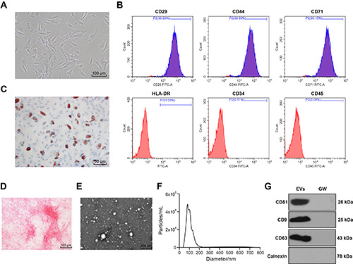
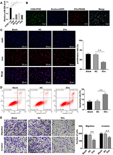
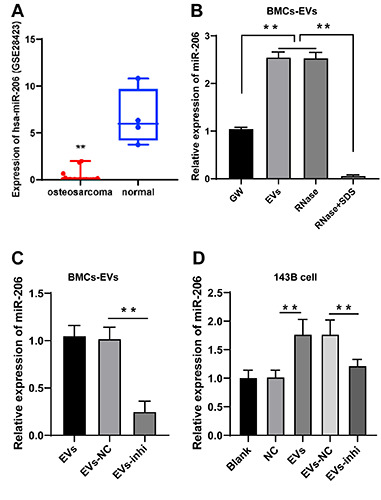
Osteosarcoma (OS) is a kind of malignant tumor originating from mesenchymal tissue Bone mesenchymal stem cells-derived extracellular vesicles (BMSCs-EVs) can play important roles in OS. This study investigated the mechanism of BMSCs-EVs on OS. BMSC surface antigens and adipogenic and osteogenic differentiation were detected by flow cytometry, and oil red O and alizarin red staining. EVs were isolated from BMSCs by differential centrifugation and identified by transmission electron microscopy, nanoparticle tracking analysis, and Western blot (WB). miR-206 and neurensin-2 (NRSN2) levels in human osteoblast hFOB 1.19 or OS cells (143B, MG-63, Saos2, HOS) were detected by RT-qPCR. Human OS cells with lower miR-206 levels were selected and treated with BMSCs-EVs or pSUPER-NRSN2. The uptake of EVs by 143B cells, cell proliferation, apoptosis, invasion, and migration were detected by immunofluorescence, 5-ethynyl-2'-deoxyuridine (EdU) and colony formation assays, flow cytometry, scratch test, and transwell assays. The binding sites between miR-206 and NRSN2 were predicted by Starbase database and verified by dual-luciferase assay. The OS xenograft model was established and treated by BMSCs-EVs. Tumor growth rate and volume, cell proliferation, and p-ERK1/2, ERK1/2, and Bcl-xL levels were detected by vernier caliper, immunohistochemistry, and WB. BMSCs-EVs were successfully extracted. miR-206 was diminished and NRSN2 was promoted in OS cells. BMSCs-EVs inhibited proliferation, migration, and invasion, and promoted apoptosis of OS cells. BMSCs-EVs carried miR-206 into OS cells. Inhibition of miR-206 in EVs partially reversed the inhibitory effect of EVs on malignant behaviors of OS cells. miR-206 targeted NRSN2. Overexpression of NRSN2 reversed the inhibitory effect of EVs on OS cells. NRSN2 activated the ERK1/2-Bcl-xL pathway. BMSC-EVs inhibited OS growth in vivo. In summary, BMSC-EVs targeted NRSN2 and inhibited the ERK1/2-Bcl-xL pathway by carrying miR-206 into OS cells, thus inhibiting OS progression.
期刊介绍:
The Journal publishes original papers concerning investigations by histochemical and immunohistochemical methods, and performed with the aid of light, super-resolution and electron microscopy, cytometry and imaging techniques. Coverage extends to:
functional cell and tissue biology in animals and plants;
cell differentiation and death;
cell-cell interaction and molecular trafficking;
biology of cell development and senescence;
nerve and muscle cell biology;
cellular basis of diseases.
The histochemical approach is nowadays essentially aimed at locating molecules in the very place where they exert their biological roles, and at describing dynamically specific chemical activities in living cells. Basic research on cell functional organization is essential for understanding the mechanisms underlying major biological processes such as differentiation, the control of tissue homeostasis, and the regulation of normal and tumor cell growth. Even more than in the past, the European Journal of Histochemistry, as a journal of functional cytology, represents the venue where cell scientists may present and discuss their original results, technical improvements and theories.

 求助内容:
求助内容: 应助结果提醒方式:


