Mark R Brinton, Russell J Stewart, Alfred K Cheung, Douglas A Christensen, Yan-Ting E Shiu
{"title":"超声波诱导的血管移植物增生轻度高热模型。","authors":"Mark R Brinton, Russell J Stewart, Alfred K Cheung, Douglas A Christensen, Yan-Ting E Shiu","doi":"10.1186/1742-4682-8-42","DOIUrl":null,"url":null,"abstract":"<p><strong>Background: </strong>Expanded polytetrafluoroethylene (ePTFE) vascular grafts frequently develop occlusive neointimal hyperplasia as a result of myofibroblast over-growth, leading to graft failure. ePTFE exhibits higher ultrasound attenuation than native soft tissues. We modelled the selective absorption of ultrasound by ePTFE, and explored the feasibility of preventing hyperplasia in ePTFE grafts by ultrasound heating. Specifically, we simulated the temperature profiles of implanted grafts and nearby soft tissues and blood under ultrasound exposure. The goal was to determine whether ultrasound exposure of an ePTFE graft can generate temperatures sufficient to prevent cell growth on the graft without damaging nearby soft tissues and blood.</p><p><strong>Methods: </strong>Ultrasound beams from two transducers (1.5 and 3.2 MHz) were simulated in two graft/tissue models, with and without an intra-graft cellular layer mimicking hyperplasia, using the finite-difference time-domain (FDTD) method. The resulting power deposition patterns were used as a heat source for the Pennes bioheat equation in a COMSOL(®) Multiphysics heat transfer model. 50°C is known to cause cell death and therefore the transducer powers were adjusted to produce a 13°C temperature rise from 37°C in the ePTFE.</p><p><strong>Results: </strong>Simulations showed that both the frequency of the transducers and the presence of hyperplasia significantly affect the power deposition patterns and subsequent temperature profiles on the grafts and nearby tissues. While neither transducer significantly raised the temperature of the blood, the 1.5-MHz transducer was less focused and heated larger volumes of the graft and nearby soft tissues than the 3.2-MHz transducer. The presence of hyperplasia had little effect on the blood's temperature, but further increased the temperature of the graft and nearby soft tissues in response to either transducer. Skin cooling and blood flow play a significant role in preventing overheating of the native tissues.</p><p><strong>Conclusions: </strong>Modelling shows that ultrasound can selectively heat ePTFE grafts and produce temperatures that cause cell death on the graft. The temperature increase in blood is negligible and that in the adjacent soft tissues may be minimized by skin cooling and using appropriate transducers. Therefore, ultrasound heating may have the potential to reduce neointimal hyperplasia and failure of ePTFE vascular grafts.</p>","PeriodicalId":51195,"journal":{"name":"Theoretical Biology and Medical Modelling","volume":" ","pages":"42"},"PeriodicalIF":0.0000,"publicationDate":"2011-11-03","publicationTypes":"Journal Article","fieldsOfStudy":null,"isOpenAccess":false,"openAccessPdf":"https://www.ncbi.nlm.nih.gov/pmc/articles/PMC3217891/pdf/","citationCount":"0","resultStr":"{\"title\":\"Modelling ultrasound-induced mild hyperthermia of hyperplasia in vascular grafts.\",\"authors\":\"Mark R Brinton, Russell J Stewart, Alfred K Cheung, Douglas A Christensen, Yan-Ting E Shiu\",\"doi\":\"10.1186/1742-4682-8-42\",\"DOIUrl\":null,\"url\":null,\"abstract\":\"<p><strong>Background: </strong>Expanded polytetrafluoroethylene (ePTFE) vascular grafts frequently develop occlusive neointimal hyperplasia as a result of myofibroblast over-growth, leading to graft failure. ePTFE exhibits higher ultrasound attenuation than native soft tissues. We modelled the selective absorption of ultrasound by ePTFE, and explored the feasibility of preventing hyperplasia in ePTFE grafts by ultrasound heating. Specifically, we simulated the temperature profiles of implanted grafts and nearby soft tissues and blood under ultrasound exposure. The goal was to determine whether ultrasound exposure of an ePTFE graft can generate temperatures sufficient to prevent cell growth on the graft without damaging nearby soft tissues and blood.</p><p><strong>Methods: </strong>Ultrasound beams from two transducers (1.5 and 3.2 MHz) were simulated in two graft/tissue models, with and without an intra-graft cellular layer mimicking hyperplasia, using the finite-difference time-domain (FDTD) method. The resulting power deposition patterns were used as a heat source for the Pennes bioheat equation in a COMSOL(®) Multiphysics heat transfer model. 50°C is known to cause cell death and therefore the transducer powers were adjusted to produce a 13°C temperature rise from 37°C in the ePTFE.</p><p><strong>Results: </strong>Simulations showed that both the frequency of the transducers and the presence of hyperplasia significantly affect the power deposition patterns and subsequent temperature profiles on the grafts and nearby tissues. While neither transducer significantly raised the temperature of the blood, the 1.5-MHz transducer was less focused and heated larger volumes of the graft and nearby soft tissues than the 3.2-MHz transducer. The presence of hyperplasia had little effect on the blood's temperature, but further increased the temperature of the graft and nearby soft tissues in response to either transducer. Skin cooling and blood flow play a significant role in preventing overheating of the native tissues.</p><p><strong>Conclusions: </strong>Modelling shows that ultrasound can selectively heat ePTFE grafts and produce temperatures that cause cell death on the graft. The temperature increase in blood is negligible and that in the adjacent soft tissues may be minimized by skin cooling and using appropriate transducers. Therefore, ultrasound heating may have the potential to reduce neointimal hyperplasia and failure of ePTFE vascular grafts.</p>\",\"PeriodicalId\":51195,\"journal\":{\"name\":\"Theoretical Biology and Medical Modelling\",\"volume\":\" \",\"pages\":\"42\"},\"PeriodicalIF\":0.0000,\"publicationDate\":\"2011-11-03\",\"publicationTypes\":\"Journal Article\",\"fieldsOfStudy\":null,\"isOpenAccess\":false,\"openAccessPdf\":\"https://www.ncbi.nlm.nih.gov/pmc/articles/PMC3217891/pdf/\",\"citationCount\":\"0\",\"resultStr\":null,\"platform\":\"Semanticscholar\",\"paperid\":null,\"PeriodicalName\":\"Theoretical Biology and Medical Modelling\",\"FirstCategoryId\":\"1085\",\"ListUrlMain\":\"https://doi.org/10.1186/1742-4682-8-42\",\"RegionNum\":0,\"RegionCategory\":null,\"ArticlePicture\":[],\"TitleCN\":null,\"AbstractTextCN\":null,\"PMCID\":null,\"EPubDate\":\"\",\"PubModel\":\"\",\"JCR\":\"Q1\",\"JCRName\":\"Mathematics\",\"Score\":null,\"Total\":0}","platform":"Semanticscholar","paperid":null,"PeriodicalName":"Theoretical Biology and Medical Modelling","FirstCategoryId":"1085","ListUrlMain":"https://doi.org/10.1186/1742-4682-8-42","RegionNum":0,"RegionCategory":null,"ArticlePicture":[],"TitleCN":null,"AbstractTextCN":null,"PMCID":null,"EPubDate":"","PubModel":"","JCR":"Q1","JCRName":"Mathematics","Score":null,"Total":0}
Modelling ultrasound-induced mild hyperthermia of hyperplasia in vascular grafts.
Background: Expanded polytetrafluoroethylene (ePTFE) vascular grafts frequently develop occlusive neointimal hyperplasia as a result of myofibroblast over-growth, leading to graft failure. ePTFE exhibits higher ultrasound attenuation than native soft tissues. We modelled the selective absorption of ultrasound by ePTFE, and explored the feasibility of preventing hyperplasia in ePTFE grafts by ultrasound heating. Specifically, we simulated the temperature profiles of implanted grafts and nearby soft tissues and blood under ultrasound exposure. The goal was to determine whether ultrasound exposure of an ePTFE graft can generate temperatures sufficient to prevent cell growth on the graft without damaging nearby soft tissues and blood.
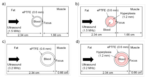
Methods: Ultrasound beams from two transducers (1.5 and 3.2 MHz) were simulated in two graft/tissue models, with and without an intra-graft cellular layer mimicking hyperplasia, using the finite-difference time-domain (FDTD) method. The resulting power deposition patterns were used as a heat source for the Pennes bioheat equation in a COMSOL(®) Multiphysics heat transfer model. 50°C is known to cause cell death and therefore the transducer powers were adjusted to produce a 13°C temperature rise from 37°C in the ePTFE.
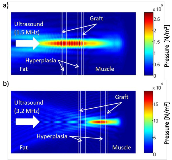
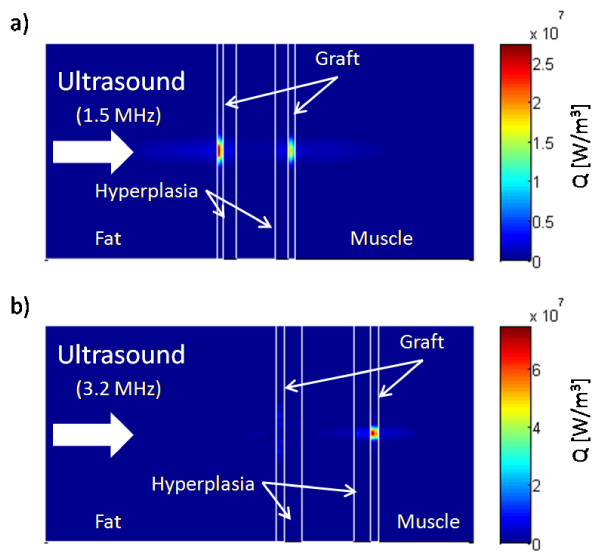
Results: Simulations showed that both the frequency of the transducers and the presence of hyperplasia significantly affect the power deposition patterns and subsequent temperature profiles on the grafts and nearby tissues. While neither transducer significantly raised the temperature of the blood, the 1.5-MHz transducer was less focused and heated larger volumes of the graft and nearby soft tissues than the 3.2-MHz transducer. The presence of hyperplasia had little effect on the blood's temperature, but further increased the temperature of the graft and nearby soft tissues in response to either transducer. Skin cooling and blood flow play a significant role in preventing overheating of the native tissues.
Conclusions: Modelling shows that ultrasound can selectively heat ePTFE grafts and produce temperatures that cause cell death on the graft. The temperature increase in blood is negligible and that in the adjacent soft tissues may be minimized by skin cooling and using appropriate transducers. Therefore, ultrasound heating may have the potential to reduce neointimal hyperplasia and failure of ePTFE vascular grafts.
期刊介绍:
Theoretical Biology and Medical Modelling is an open access peer-reviewed journal adopting a broad definition of "biology" and focusing on theoretical ideas and models associated with developments in biology and medicine. Mathematicians, biologists and clinicians of various specialisms, philosophers and historians of science are all contributing to the emergence of novel concepts in an age of systems biology, bioinformatics and computer modelling. This is the field in which Theoretical Biology and Medical Modelling operates. We welcome submissions that are technically sound and offering either improved understanding in biology and medicine or progress in theory or method.
 求助内容:
求助内容: 应助结果提醒方式:


