{"title":"x连锁Alport综合征斑视网膜和颞黄斑变薄的电生理评价。","authors":"Adel Al Akeely, Patrik Schatz, Amro Alhazimi","doi":"10.4103/meajo.meajo_7_21","DOIUrl":null,"url":null,"abstract":"<p><p>We report a 39-year-old with Alport's syndrome. The patient presented with anterior lenticonus, cataract, and a corrected distance visual acuity of 20/25 and 20/60 in the right and left eyes, respectively. Fundus examination revealed generalized retinal flecks sparing the fovea in both eyes. Optical coherence topography showed temporal macular thinning. Normal fundus autofluorescence was observed in both eyes. Full-field electroretinography (ERG) demonstrated normal photopic and scotopic responses, while multifocal ERG showed no reduction of amplitudes generated from the temporal thinned macula, compared to the nasal macula, indicating preserved functional integrity of the retina.</p>","PeriodicalId":18740,"journal":{"name":"Middle East African Journal of Ophthalmology","volume":"28 4","pages":"257-259"},"PeriodicalIF":0.3000,"publicationDate":"2022-04-30","publicationTypes":"Journal Article","fieldsOfStudy":null,"isOpenAccess":false,"openAccessPdf":"https://www.ncbi.nlm.nih.gov/pmc/articles/PMC9198527/pdf/","citationCount":"0","resultStr":"{\"title\":\"Electrophysiological Evaluation of Fleck Retina and Temporal Macular Thinning in X-Linked Alport's Syndrome.\",\"authors\":\"Adel Al Akeely, Patrik Schatz, Amro Alhazimi\",\"doi\":\"10.4103/meajo.meajo_7_21\",\"DOIUrl\":null,\"url\":null,\"abstract\":\"<p><p>We report a 39-year-old with Alport's syndrome. The patient presented with anterior lenticonus, cataract, and a corrected distance visual acuity of 20/25 and 20/60 in the right and left eyes, respectively. Fundus examination revealed generalized retinal flecks sparing the fovea in both eyes. Optical coherence topography showed temporal macular thinning. Normal fundus autofluorescence was observed in both eyes. Full-field electroretinography (ERG) demonstrated normal photopic and scotopic responses, while multifocal ERG showed no reduction of amplitudes generated from the temporal thinned macula, compared to the nasal macula, indicating preserved functional integrity of the retina.</p>\",\"PeriodicalId\":18740,\"journal\":{\"name\":\"Middle East African Journal of Ophthalmology\",\"volume\":\"28 4\",\"pages\":\"257-259\"},\"PeriodicalIF\":0.3000,\"publicationDate\":\"2022-04-30\",\"publicationTypes\":\"Journal Article\",\"fieldsOfStudy\":null,\"isOpenAccess\":false,\"openAccessPdf\":\"https://www.ncbi.nlm.nih.gov/pmc/articles/PMC9198527/pdf/\",\"citationCount\":\"0\",\"resultStr\":null,\"platform\":\"Semanticscholar\",\"paperid\":null,\"PeriodicalName\":\"Middle East African Journal of Ophthalmology\",\"FirstCategoryId\":\"1085\",\"ListUrlMain\":\"https://doi.org/10.4103/meajo.meajo_7_21\",\"RegionNum\":0,\"RegionCategory\":null,\"ArticlePicture\":[],\"TitleCN\":null,\"AbstractTextCN\":null,\"PMCID\":null,\"EPubDate\":\"2021/10/1 0:00:00\",\"PubModel\":\"eCollection\",\"JCR\":\"Q4\",\"JCRName\":\"OPHTHALMOLOGY\",\"Score\":null,\"Total\":0}","platform":"Semanticscholar","paperid":null,"PeriodicalName":"Middle East African Journal of Ophthalmology","FirstCategoryId":"1085","ListUrlMain":"https://doi.org/10.4103/meajo.meajo_7_21","RegionNum":0,"RegionCategory":null,"ArticlePicture":[],"TitleCN":null,"AbstractTextCN":null,"PMCID":null,"EPubDate":"2021/10/1 0:00:00","PubModel":"eCollection","JCR":"Q4","JCRName":"OPHTHALMOLOGY","Score":null,"Total":0}
Electrophysiological Evaluation of Fleck Retina and Temporal Macular Thinning in X-Linked Alport's Syndrome.
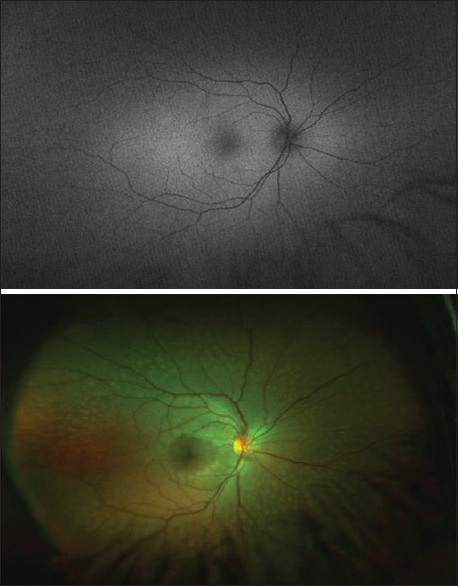
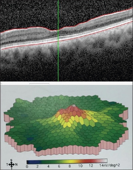
We report a 39-year-old with Alport's syndrome. The patient presented with anterior lenticonus, cataract, and a corrected distance visual acuity of 20/25 and 20/60 in the right and left eyes, respectively. Fundus examination revealed generalized retinal flecks sparing the fovea in both eyes. Optical coherence topography showed temporal macular thinning. Normal fundus autofluorescence was observed in both eyes. Full-field electroretinography (ERG) demonstrated normal photopic and scotopic responses, while multifocal ERG showed no reduction of amplitudes generated from the temporal thinned macula, compared to the nasal macula, indicating preserved functional integrity of the retina.
期刊介绍:
The Middle East African Journal of Ophthalmology (MEAJO), published four times per year in print and online, is an official journal of the Middle East African Council of Ophthalmology (MEACO). It is an international, peer-reviewed journal whose mission includes publication of original research of interest to ophthalmologists in the Middle East and Africa, and to provide readers with high quality educational review articles from world-renown experts. MEAJO, previously known as Middle East Journal of Ophthalmology (MEJO) was founded by Dr Akef El Maghraby in 1993.

 求助内容:
求助内容: 应助结果提醒方式:


