Bin Han, Hui Zhao, Xingji Gong, Jinping Sun, Song Chi, Tao Liu, Anmu Xie
{"title":"PGC-1α上调可减轻氧糖剥夺诱导的海马神经元损伤。","authors":"Bin Han, Hui Zhao, Xingji Gong, Jinping Sun, Song Chi, Tao Liu, Anmu Xie","doi":"10.1155/2022/9682999","DOIUrl":null,"url":null,"abstract":"<p><p>Hippocampal neuronal damage likely underlies cognitive impairment in vascular dementia (VaD). PPAR<i>γ</i> coactivator-1<i>α</i> (PGC-1<i>α</i>) is a master regulator of mitochondrial biogenesis. However, the role and the precise mechanism of how PGC-1<i>α</i> alleviates hippocampal neuronal injury remain unknown. To address this question, HT-22 cells, an immortalized hippocampal neuron cell line, with or without PGC-1<i>α</i> overexpression were subjected to oxygen-glucose deprivation (OGD), which mimics the circumstance of chronic cerebral hypoperfusion in VaD. After OGD, cell viability was assessed using the MTS assay. The mitochondrial function and reactive oxygen species (ROS) were both detected. ChIP-Seq analysis was employed to discover the underlying molecular mechanism of PGC-1<i>α</i>-mediated neuroprotective effects. Our results showed that mitochondrial membrane potentials were increased and ROS production was decreased in PGC-1<i>α</i> overexpressing cells, which increased cell viability. The further bioinformatics analysis from ChIP-Seq data indicated that PGC-1<i>α</i> may participate in the regulation of apoptosis, autophagy, and mitophagy pathways in HT-22 cells. We found that PGC-1<i>α</i> promoted the LC3-II formation and reduced the neuronal apoptosis determined by TUNEL staining. In addition, PGC-1<i>α</i> upregulated the expressions of mitochondrial antioxidants, including SOD2, Trx2, and Prx3. In summary, our findings indicate that PGC-1<i>α</i> may attenuate OGD-induced hippocampal neuronal damage by regulating multiple mechanisms, like autophagy and mitochondrial function. Thus, PGC-1<i>α</i> may be a potential therapeutic target for hippocampal damage associated with cognitive impairment.</p>","PeriodicalId":51299,"journal":{"name":"Neural Plasticity","volume":" ","pages":"9682999"},"PeriodicalIF":3.7000,"publicationDate":"2022-06-09","publicationTypes":"Journal Article","fieldsOfStudy":null,"isOpenAccess":false,"openAccessPdf":"https://www.ncbi.nlm.nih.gov/pmc/articles/PMC9203239/pdf/","citationCount":"3","resultStr":"{\"title\":\"Upregulation of PGC-1<i>α</i> Attenuates Oxygen-Glucose Deprivation-Induced Hippocampal Neuronal Injury.\",\"authors\":\"Bin Han, Hui Zhao, Xingji Gong, Jinping Sun, Song Chi, Tao Liu, Anmu Xie\",\"doi\":\"10.1155/2022/9682999\",\"DOIUrl\":null,\"url\":null,\"abstract\":\"<p><p>Hippocampal neuronal damage likely underlies cognitive impairment in vascular dementia (VaD). PPAR<i>γ</i> coactivator-1<i>α</i> (PGC-1<i>α</i>) is a master regulator of mitochondrial biogenesis. However, the role and the precise mechanism of how PGC-1<i>α</i> alleviates hippocampal neuronal injury remain unknown. To address this question, HT-22 cells, an immortalized hippocampal neuron cell line, with or without PGC-1<i>α</i> overexpression were subjected to oxygen-glucose deprivation (OGD), which mimics the circumstance of chronic cerebral hypoperfusion in VaD. After OGD, cell viability was assessed using the MTS assay. The mitochondrial function and reactive oxygen species (ROS) were both detected. ChIP-Seq analysis was employed to discover the underlying molecular mechanism of PGC-1<i>α</i>-mediated neuroprotective effects. Our results showed that mitochondrial membrane potentials were increased and ROS production was decreased in PGC-1<i>α</i> overexpressing cells, which increased cell viability. The further bioinformatics analysis from ChIP-Seq data indicated that PGC-1<i>α</i> may participate in the regulation of apoptosis, autophagy, and mitophagy pathways in HT-22 cells. We found that PGC-1<i>α</i> promoted the LC3-II formation and reduced the neuronal apoptosis determined by TUNEL staining. In addition, PGC-1<i>α</i> upregulated the expressions of mitochondrial antioxidants, including SOD2, Trx2, and Prx3. In summary, our findings indicate that PGC-1<i>α</i> may attenuate OGD-induced hippocampal neuronal damage by regulating multiple mechanisms, like autophagy and mitochondrial function. Thus, PGC-1<i>α</i> may be a potential therapeutic target for hippocampal damage associated with cognitive impairment.</p>\",\"PeriodicalId\":51299,\"journal\":{\"name\":\"Neural Plasticity\",\"volume\":\" \",\"pages\":\"9682999\"},\"PeriodicalIF\":3.7000,\"publicationDate\":\"2022-06-09\",\"publicationTypes\":\"Journal Article\",\"fieldsOfStudy\":null,\"isOpenAccess\":false,\"openAccessPdf\":\"https://www.ncbi.nlm.nih.gov/pmc/articles/PMC9203239/pdf/\",\"citationCount\":\"3\",\"resultStr\":null,\"platform\":\"Semanticscholar\",\"paperid\":null,\"PeriodicalName\":\"Neural Plasticity\",\"FirstCategoryId\":\"3\",\"ListUrlMain\":\"https://doi.org/10.1155/2022/9682999\",\"RegionNum\":4,\"RegionCategory\":\"医学\",\"ArticlePicture\":[],\"TitleCN\":null,\"AbstractTextCN\":null,\"PMCID\":null,\"EPubDate\":\"2022/1/1 0:00:00\",\"PubModel\":\"eCollection\",\"JCR\":\"Q2\",\"JCRName\":\"NEUROSCIENCES\",\"Score\":null,\"Total\":0}","platform":"Semanticscholar","paperid":null,"PeriodicalName":"Neural Plasticity","FirstCategoryId":"3","ListUrlMain":"https://doi.org/10.1155/2022/9682999","RegionNum":4,"RegionCategory":"医学","ArticlePicture":[],"TitleCN":null,"AbstractTextCN":null,"PMCID":null,"EPubDate":"2022/1/1 0:00:00","PubModel":"eCollection","JCR":"Q2","JCRName":"NEUROSCIENCES","Score":null,"Total":0}
Upregulation of PGC-1α Attenuates Oxygen-Glucose Deprivation-Induced Hippocampal Neuronal Injury.
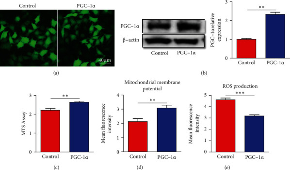
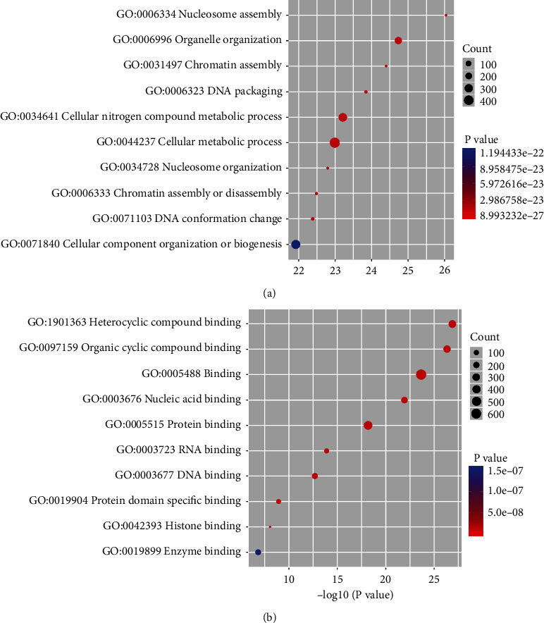
Hippocampal neuronal damage likely underlies cognitive impairment in vascular dementia (VaD). PPARγ coactivator-1α (PGC-1α) is a master regulator of mitochondrial biogenesis. However, the role and the precise mechanism of how PGC-1α alleviates hippocampal neuronal injury remain unknown. To address this question, HT-22 cells, an immortalized hippocampal neuron cell line, with or without PGC-1α overexpression were subjected to oxygen-glucose deprivation (OGD), which mimics the circumstance of chronic cerebral hypoperfusion in VaD. After OGD, cell viability was assessed using the MTS assay. The mitochondrial function and reactive oxygen species (ROS) were both detected. ChIP-Seq analysis was employed to discover the underlying molecular mechanism of PGC-1α-mediated neuroprotective effects. Our results showed that mitochondrial membrane potentials were increased and ROS production was decreased in PGC-1α overexpressing cells, which increased cell viability. The further bioinformatics analysis from ChIP-Seq data indicated that PGC-1α may participate in the regulation of apoptosis, autophagy, and mitophagy pathways in HT-22 cells. We found that PGC-1α promoted the LC3-II formation and reduced the neuronal apoptosis determined by TUNEL staining. In addition, PGC-1α upregulated the expressions of mitochondrial antioxidants, including SOD2, Trx2, and Prx3. In summary, our findings indicate that PGC-1α may attenuate OGD-induced hippocampal neuronal damage by regulating multiple mechanisms, like autophagy and mitochondrial function. Thus, PGC-1α may be a potential therapeutic target for hippocampal damage associated with cognitive impairment.
期刊介绍:
Neural Plasticity is an international, interdisciplinary journal dedicated to the publication of articles related to all aspects of neural plasticity, with special emphasis on its functional significance as reflected in behavior and in psychopathology. Neural Plasticity publishes research and review articles from the entire range of relevant disciplines, including basic neuroscience, behavioral neuroscience, cognitive neuroscience, biological psychology, and biological psychiatry.

 求助内容:
求助内容: 应助结果提醒方式:


