Yongwei Lin, Zhipeng Zhou, Lang Xie, Yongsheng Huang, Zhenghua Qiu, Lili Ye, Chunhui Cui
{"title":"miR-939和miR-376A通过诱骗策略抑制NF-κB和NFAT表达对溃疡性结肠炎的影响","authors":"Yongwei Lin, Zhipeng Zhou, Lang Xie, Yongsheng Huang, Zhenghua Qiu, Lili Ye, Chunhui Cui","doi":"10.4081/ejh.2022.3316","DOIUrl":null,"url":null,"abstract":"<p><p>The aim of this study was to explore the effects of miR-939 and miR-376A on the pathogenesis of ulcerative colitis (UC) by using a decoy strategy to regulate the expression of nuclear transcription factor kappa B (NF-κB) and nuclear factor of activated T cells (NFAT). Such strategies represent a potential novel treatment for UC. Quantitative polymerase chain reaction (qPCR) analysis was used to detect the differences between the expression of miR-939, miR-376a, NF-κB, NFAT in the tissue samples from the resting and active stages of UC and healthy controls, and analyzed the correlation. The electrophoretic mobility shift assay was used to validate the ability of miRNAs to bind to NF-κB and NFAT. The expression of components of the intestinal barrier in UC and changes in apoptosis-related factors were examined by western blotting, qPCR, and immunofluorescence. After a dextran sulfate sodium (DSS)-induced mouse model of UC was established, the morphological changes in the colonic tissues of mice, the changes in serum inflammatory factors, and the changes in urine protein or urine leukocytes, liver enzymes, and prothrombin time were measured to examine intestinal permeability. The expression of miR-939 and miR-376a in human UC tissue was significantly lower than that in the normal control tissue, and was negatively correlated with the expression of NF-κB and NFAT. miR-939 and miR-376a decoy strategies resulted in a beneficial increase in the expression of claudins, occludins, and ZO-1 protein and inhibited apoptosis in intestinal epithelial cells. The disease activity index of the UC model group was significantly higher than that of the normal control group. The expression of inflammatory factors in the decoy group was higher than that in the UC model group. Therefore, from the experimental results, it can be concluded that using miR-939 and miR-376a to trap NF-κB and NFAT inhibits the activation of transcription factors NF-κB and NFAT, which in turn inhibits the expression of inflammatory factors and results in partial recovery of the intestinal barrier in UC. The decoy strategy inhibited apoptosis in the target cells and had a therapeutic effect in the mice model of UC. This study provides new ideas for the development of future clinical therapies for UC.</p>","PeriodicalId":50487,"journal":{"name":"European Journal of Histochemistry","volume":"66 1","pages":""},"PeriodicalIF":2.1000,"publicationDate":"2022-02-15","publicationTypes":"Journal Article","fieldsOfStudy":null,"isOpenAccess":false,"openAccessPdf":"https://ftp.ncbi.nlm.nih.gov/pub/pmc/oa_pdf/21/1f/ejh-66-1-3316.PMC8875791.pdf","citationCount":"2","resultStr":"{\"title\":\"Effects of miR-939 and miR-376A on ulcerative colitis using a decoy strategy to inhibit NF-κB and NFAT expression.\",\"authors\":\"Yongwei Lin, Zhipeng Zhou, Lang Xie, Yongsheng Huang, Zhenghua Qiu, Lili Ye, Chunhui Cui\",\"doi\":\"10.4081/ejh.2022.3316\",\"DOIUrl\":null,\"url\":null,\"abstract\":\"<p><p>The aim of this study was to explore the effects of miR-939 and miR-376A on the pathogenesis of ulcerative colitis (UC) by using a decoy strategy to regulate the expression of nuclear transcription factor kappa B (NF-κB) and nuclear factor of activated T cells (NFAT). Such strategies represent a potential novel treatment for UC. Quantitative polymerase chain reaction (qPCR) analysis was used to detect the differences between the expression of miR-939, miR-376a, NF-κB, NFAT in the tissue samples from the resting and active stages of UC and healthy controls, and analyzed the correlation. The electrophoretic mobility shift assay was used to validate the ability of miRNAs to bind to NF-κB and NFAT. The expression of components of the intestinal barrier in UC and changes in apoptosis-related factors were examined by western blotting, qPCR, and immunofluorescence. After a dextran sulfate sodium (DSS)-induced mouse model of UC was established, the morphological changes in the colonic tissues of mice, the changes in serum inflammatory factors, and the changes in urine protein or urine leukocytes, liver enzymes, and prothrombin time were measured to examine intestinal permeability. The expression of miR-939 and miR-376a in human UC tissue was significantly lower than that in the normal control tissue, and was negatively correlated with the expression of NF-κB and NFAT. miR-939 and miR-376a decoy strategies resulted in a beneficial increase in the expression of claudins, occludins, and ZO-1 protein and inhibited apoptosis in intestinal epithelial cells. The disease activity index of the UC model group was significantly higher than that of the normal control group. The expression of inflammatory factors in the decoy group was higher than that in the UC model group. Therefore, from the experimental results, it can be concluded that using miR-939 and miR-376a to trap NF-κB and NFAT inhibits the activation of transcription factors NF-κB and NFAT, which in turn inhibits the expression of inflammatory factors and results in partial recovery of the intestinal barrier in UC. The decoy strategy inhibited apoptosis in the target cells and had a therapeutic effect in the mice model of UC. This study provides new ideas for the development of future clinical therapies for UC.</p>\",\"PeriodicalId\":50487,\"journal\":{\"name\":\"European Journal of Histochemistry\",\"volume\":\"66 1\",\"pages\":\"\"},\"PeriodicalIF\":2.1000,\"publicationDate\":\"2022-02-15\",\"publicationTypes\":\"Journal Article\",\"fieldsOfStudy\":null,\"isOpenAccess\":false,\"openAccessPdf\":\"https://ftp.ncbi.nlm.nih.gov/pub/pmc/oa_pdf/21/1f/ejh-66-1-3316.PMC8875791.pdf\",\"citationCount\":\"2\",\"resultStr\":null,\"platform\":\"Semanticscholar\",\"paperid\":null,\"PeriodicalName\":\"European Journal of Histochemistry\",\"FirstCategoryId\":\"99\",\"ListUrlMain\":\"https://doi.org/10.4081/ejh.2022.3316\",\"RegionNum\":4,\"RegionCategory\":\"生物学\",\"ArticlePicture\":[],\"TitleCN\":null,\"AbstractTextCN\":null,\"PMCID\":null,\"EPubDate\":\"\",\"PubModel\":\"\",\"JCR\":\"Q4\",\"JCRName\":\"CELL BIOLOGY\",\"Score\":null,\"Total\":0}","platform":"Semanticscholar","paperid":null,"PeriodicalName":"European Journal of Histochemistry","FirstCategoryId":"99","ListUrlMain":"https://doi.org/10.4081/ejh.2022.3316","RegionNum":4,"RegionCategory":"生物学","ArticlePicture":[],"TitleCN":null,"AbstractTextCN":null,"PMCID":null,"EPubDate":"","PubModel":"","JCR":"Q4","JCRName":"CELL BIOLOGY","Score":null,"Total":0}
Effects of miR-939 and miR-376A on ulcerative colitis using a decoy strategy to inhibit NF-κB and NFAT expression.
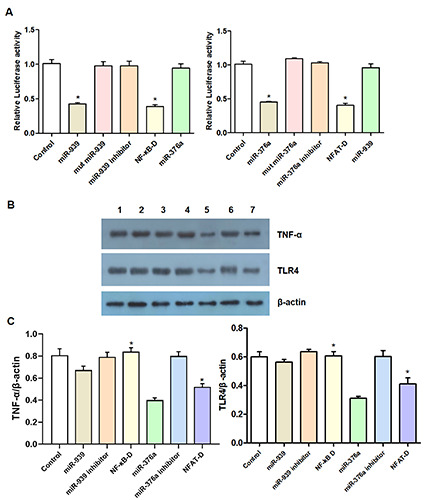
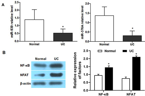
The aim of this study was to explore the effects of miR-939 and miR-376A on the pathogenesis of ulcerative colitis (UC) by using a decoy strategy to regulate the expression of nuclear transcription factor kappa B (NF-κB) and nuclear factor of activated T cells (NFAT). Such strategies represent a potential novel treatment for UC. Quantitative polymerase chain reaction (qPCR) analysis was used to detect the differences between the expression of miR-939, miR-376a, NF-κB, NFAT in the tissue samples from the resting and active stages of UC and healthy controls, and analyzed the correlation. The electrophoretic mobility shift assay was used to validate the ability of miRNAs to bind to NF-κB and NFAT. The expression of components of the intestinal barrier in UC and changes in apoptosis-related factors were examined by western blotting, qPCR, and immunofluorescence. After a dextran sulfate sodium (DSS)-induced mouse model of UC was established, the morphological changes in the colonic tissues of mice, the changes in serum inflammatory factors, and the changes in urine protein or urine leukocytes, liver enzymes, and prothrombin time were measured to examine intestinal permeability. The expression of miR-939 and miR-376a in human UC tissue was significantly lower than that in the normal control tissue, and was negatively correlated with the expression of NF-κB and NFAT. miR-939 and miR-376a decoy strategies resulted in a beneficial increase in the expression of claudins, occludins, and ZO-1 protein and inhibited apoptosis in intestinal epithelial cells. The disease activity index of the UC model group was significantly higher than that of the normal control group. The expression of inflammatory factors in the decoy group was higher than that in the UC model group. Therefore, from the experimental results, it can be concluded that using miR-939 and miR-376a to trap NF-κB and NFAT inhibits the activation of transcription factors NF-κB and NFAT, which in turn inhibits the expression of inflammatory factors and results in partial recovery of the intestinal barrier in UC. The decoy strategy inhibited apoptosis in the target cells and had a therapeutic effect in the mice model of UC. This study provides new ideas for the development of future clinical therapies for UC.
期刊介绍:
The Journal publishes original papers concerning investigations by histochemical and immunohistochemical methods, and performed with the aid of light, super-resolution and electron microscopy, cytometry and imaging techniques. Coverage extends to:
functional cell and tissue biology in animals and plants;
cell differentiation and death;
cell-cell interaction and molecular trafficking;
biology of cell development and senescence;
nerve and muscle cell biology;
cellular basis of diseases.
The histochemical approach is nowadays essentially aimed at locating molecules in the very place where they exert their biological roles, and at describing dynamically specific chemical activities in living cells. Basic research on cell functional organization is essential for understanding the mechanisms underlying major biological processes such as differentiation, the control of tissue homeostasis, and the regulation of normal and tumor cell growth. Even more than in the past, the European Journal of Histochemistry, as a journal of functional cytology, represents the venue where cell scientists may present and discuss their original results, technical improvements and theories.

 求助内容:
求助内容: 应助结果提醒方式:


