A V Krauze, Y Zhuge, R Zhao, E Tasci, K Camphausen
{"title":"人工智能驱动的中枢神经系统肿瘤图像分析--传统机器学习、深度学习和混合模型。","authors":"A V Krauze, Y Zhuge, R Zhao, E Tasci, K Camphausen","doi":"10.26502/jbb.2642-91280046","DOIUrl":null,"url":null,"abstract":"<p><p>The interpretation of imaging in medicine in general and in oncology specifically remains problematic due to several limitations which include the need to incorporate detailed clinical history, patient and disease-specific history, clinical exam features, previous and ongoing treatment, and account for the dependency on reproducible human interpretation of multiple factors with incomplete data linkage. To standardize reporting, minimize bias, expedite management, and improve outcomes, the use of Artificial Intelligence (AI) has gained significant prominence in imaging analysis. In oncology, AI methods have as a result been explored in most cancer types with ongoing progress in employing AI towards imaging for oncology treatment, assessing treatment response, and understanding and communicating prognosis. Challenges remain with limited available data sets, variability in imaging changes over time augmented by a growing heterogeneity in analysis approaches. We review the imaging analysis workflow and examine how hand-crafted features also referred to as traditional Machine Learning (ML), Deep Learning (DL) approaches, and hybrid analyses, are being employed in AI-driven imaging analysis in central nervous system tumors. ML, DL, and hybrid approaches coexist, and their combination may produce superior results although data in this space is as yet novel, and conclusions and pitfalls have yet to be fully explored. We note the growing technical complexities that may become increasingly separated from the clinic and enforce the acute need for clinician engagement to guide progress and ensure that conclusions derived from AI-driven imaging analysis reflect that same level of scrutiny lent to other avenues of clinical research.</p>","PeriodicalId":15066,"journal":{"name":"Journal of Biotechnology and Biomedicine","volume":"5 1","pages":"1-19"},"PeriodicalIF":0.0000,"publicationDate":"2022-01-01","publicationTypes":"Journal Article","fieldsOfStudy":null,"isOpenAccess":false,"openAccessPdf":"https://www.ncbi.nlm.nih.gov/pmc/articles/PMC8802234/pdf/","citationCount":"0","resultStr":"{\"title\":\"AI-Driven Image Analysis in Central Nervous System Tumors-Traditional Machine Learning, Deep Learning and Hybrid Models.\",\"authors\":\"A V Krauze, Y Zhuge, R Zhao, E Tasci, K Camphausen\",\"doi\":\"10.26502/jbb.2642-91280046\",\"DOIUrl\":null,\"url\":null,\"abstract\":\"<p><p>The interpretation of imaging in medicine in general and in oncology specifically remains problematic due to several limitations which include the need to incorporate detailed clinical history, patient and disease-specific history, clinical exam features, previous and ongoing treatment, and account for the dependency on reproducible human interpretation of multiple factors with incomplete data linkage. To standardize reporting, minimize bias, expedite management, and improve outcomes, the use of Artificial Intelligence (AI) has gained significant prominence in imaging analysis. In oncology, AI methods have as a result been explored in most cancer types with ongoing progress in employing AI towards imaging for oncology treatment, assessing treatment response, and understanding and communicating prognosis. Challenges remain with limited available data sets, variability in imaging changes over time augmented by a growing heterogeneity in analysis approaches. We review the imaging analysis workflow and examine how hand-crafted features also referred to as traditional Machine Learning (ML), Deep Learning (DL) approaches, and hybrid analyses, are being employed in AI-driven imaging analysis in central nervous system tumors. ML, DL, and hybrid approaches coexist, and their combination may produce superior results although data in this space is as yet novel, and conclusions and pitfalls have yet to be fully explored. We note the growing technical complexities that may become increasingly separated from the clinic and enforce the acute need for clinician engagement to guide progress and ensure that conclusions derived from AI-driven imaging analysis reflect that same level of scrutiny lent to other avenues of clinical research.</p>\",\"PeriodicalId\":15066,\"journal\":{\"name\":\"Journal of Biotechnology and Biomedicine\",\"volume\":\"5 1\",\"pages\":\"1-19\"},\"PeriodicalIF\":0.0000,\"publicationDate\":\"2022-01-01\",\"publicationTypes\":\"Journal Article\",\"fieldsOfStudy\":null,\"isOpenAccess\":false,\"openAccessPdf\":\"https://www.ncbi.nlm.nih.gov/pmc/articles/PMC8802234/pdf/\",\"citationCount\":\"0\",\"resultStr\":null,\"platform\":\"Semanticscholar\",\"paperid\":null,\"PeriodicalName\":\"Journal of Biotechnology and Biomedicine\",\"FirstCategoryId\":\"1085\",\"ListUrlMain\":\"https://doi.org/10.26502/jbb.2642-91280046\",\"RegionNum\":0,\"RegionCategory\":null,\"ArticlePicture\":[],\"TitleCN\":null,\"AbstractTextCN\":null,\"PMCID\":null,\"EPubDate\":\"2022/1/10 0:00:00\",\"PubModel\":\"Epub\",\"JCR\":\"\",\"JCRName\":\"\",\"Score\":null,\"Total\":0}","platform":"Semanticscholar","paperid":null,"PeriodicalName":"Journal of Biotechnology and Biomedicine","FirstCategoryId":"1085","ListUrlMain":"https://doi.org/10.26502/jbb.2642-91280046","RegionNum":0,"RegionCategory":null,"ArticlePicture":[],"TitleCN":null,"AbstractTextCN":null,"PMCID":null,"EPubDate":"2022/1/10 0:00:00","PubModel":"Epub","JCR":"","JCRName":"","Score":null,"Total":0}
AI-Driven Image Analysis in Central Nervous System Tumors-Traditional Machine Learning, Deep Learning and Hybrid Models.
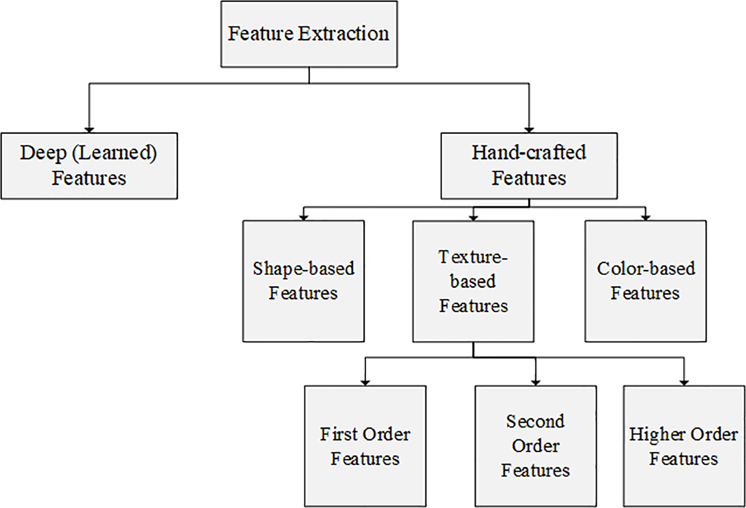
The interpretation of imaging in medicine in general and in oncology specifically remains problematic due to several limitations which include the need to incorporate detailed clinical history, patient and disease-specific history, clinical exam features, previous and ongoing treatment, and account for the dependency on reproducible human interpretation of multiple factors with incomplete data linkage. To standardize reporting, minimize bias, expedite management, and improve outcomes, the use of Artificial Intelligence (AI) has gained significant prominence in imaging analysis. In oncology, AI methods have as a result been explored in most cancer types with ongoing progress in employing AI towards imaging for oncology treatment, assessing treatment response, and understanding and communicating prognosis. Challenges remain with limited available data sets, variability in imaging changes over time augmented by a growing heterogeneity in analysis approaches. We review the imaging analysis workflow and examine how hand-crafted features also referred to as traditional Machine Learning (ML), Deep Learning (DL) approaches, and hybrid analyses, are being employed in AI-driven imaging analysis in central nervous system tumors. ML, DL, and hybrid approaches coexist, and their combination may produce superior results although data in this space is as yet novel, and conclusions and pitfalls have yet to be fully explored. We note the growing technical complexities that may become increasingly separated from the clinic and enforce the acute need for clinician engagement to guide progress and ensure that conclusions derived from AI-driven imaging analysis reflect that same level of scrutiny lent to other avenues of clinical research.


 求助内容:
求助内容: 应助结果提醒方式:


