{"title":"18F-FDG、18f -氟乙酸酯和18F-FEPPA在胆管结扎大鼠模型中肝纤维化成像的比较","authors":"Chun-Yi Wu, Hsin-Hua Hsieh, Pei-An Chu, Wen-Hsiang Hong, Ting-Yu Chang, Chia-Fang Hsu, Siao-Ting Lin, Po-Hsun Su, Shin-Lei Peng","doi":"10.1155/2021/7545284","DOIUrl":null,"url":null,"abstract":"<p><p>Developing sensitive diagnostic methods for a longitudinal evaluation of the status of liver fibrosis is a priority. This study is aimed at assessing the significance of longitudinal positron emission tomography (PET) imaging with <sup>18</sup>F-labeling tracers for assessing liver fibrosis in a rat model with bile duct ligation (BDL). Twenty-one 6-week-old Sprague-Dawley male rats were used in this study. Longitudinal PET images using [<sup>18</sup>F]N-2-(2-fluoroethoxy)benzyl)-N-(4-phenoxypyridin-3-yl)acetamide ([<sup>18</sup>F]FEPPA) (<i>n</i> = 3), [<sup>18</sup>F]fluoroacetate ([<sup>18</sup>F]FAc) (<i>n</i> = 3), and 18F-fluoro-2-deoxy-D-glucose ([<sup>18</sup>F]FDG) (<i>n</i> = 3) were obtained at 0, 1, and 2 weeks after BDL. Biochemical assays, histological assays, immunohistochemical staining assays, and next generation sequencing analyses were also performed at 0 (<i>n</i> = 3), 1 (<i>n</i> = 3), 2 (<i>n</i> = 3), and 3 (<i>n</i> = 3) weeks after BDL, which demonstrated the severe damage in rat livers after BDL. Regarding [<sup>18</sup>F]FEPPA and [<sup>18</sup>F]FDG, there was a significantly higher uptake in the liver after BDL (both <i>P</i> < 0.05), which lasted until week 2. However, the uptake of [<sup>18</sup>F]FAc in the liver was not significantly different before and after BDL (<i>P</i> = 0.28). Collectively, both [<sup>18</sup>F]FEPPA and [<sup>18</sup>F]FDG can serve as sensitive probes for detecting the liver fibrosis. However, [<sup>18</sup>F]FAc is not recommended to diagnose liver fibrosis.</p>","PeriodicalId":18855,"journal":{"name":"Molecular Imaging","volume":" ","pages":"7545284"},"PeriodicalIF":2.4000,"publicationDate":"2021-11-27","publicationTypes":"Journal Article","fieldsOfStudy":null,"isOpenAccess":false,"openAccessPdf":"https://www.ncbi.nlm.nih.gov/pmc/articles/PMC8654319/pdf/","citationCount":"2","resultStr":"{\"title\":\"Comparison of <sup>18</sup>F-FDG, <sup>18</sup>F-Fluoroacetate, and <sup>18</sup>F-FEPPA for Imaging Liver Fibrosis in a Bile Duct-Ligated Rat Model.\",\"authors\":\"Chun-Yi Wu, Hsin-Hua Hsieh, Pei-An Chu, Wen-Hsiang Hong, Ting-Yu Chang, Chia-Fang Hsu, Siao-Ting Lin, Po-Hsun Su, Shin-Lei Peng\",\"doi\":\"10.1155/2021/7545284\",\"DOIUrl\":null,\"url\":null,\"abstract\":\"<p><p>Developing sensitive diagnostic methods for a longitudinal evaluation of the status of liver fibrosis is a priority. This study is aimed at assessing the significance of longitudinal positron emission tomography (PET) imaging with <sup>18</sup>F-labeling tracers for assessing liver fibrosis in a rat model with bile duct ligation (BDL). Twenty-one 6-week-old Sprague-Dawley male rats were used in this study. Longitudinal PET images using [<sup>18</sup>F]N-2-(2-fluoroethoxy)benzyl)-N-(4-phenoxypyridin-3-yl)acetamide ([<sup>18</sup>F]FEPPA) (<i>n</i> = 3), [<sup>18</sup>F]fluoroacetate ([<sup>18</sup>F]FAc) (<i>n</i> = 3), and 18F-fluoro-2-deoxy-D-glucose ([<sup>18</sup>F]FDG) (<i>n</i> = 3) were obtained at 0, 1, and 2 weeks after BDL. Biochemical assays, histological assays, immunohistochemical staining assays, and next generation sequencing analyses were also performed at 0 (<i>n</i> = 3), 1 (<i>n</i> = 3), 2 (<i>n</i> = 3), and 3 (<i>n</i> = 3) weeks after BDL, which demonstrated the severe damage in rat livers after BDL. Regarding [<sup>18</sup>F]FEPPA and [<sup>18</sup>F]FDG, there was a significantly higher uptake in the liver after BDL (both <i>P</i> < 0.05), which lasted until week 2. However, the uptake of [<sup>18</sup>F]FAc in the liver was not significantly different before and after BDL (<i>P</i> = 0.28). Collectively, both [<sup>18</sup>F]FEPPA and [<sup>18</sup>F]FDG can serve as sensitive probes for detecting the liver fibrosis. However, [<sup>18</sup>F]FAc is not recommended to diagnose liver fibrosis.</p>\",\"PeriodicalId\":18855,\"journal\":{\"name\":\"Molecular Imaging\",\"volume\":\" \",\"pages\":\"7545284\"},\"PeriodicalIF\":2.4000,\"publicationDate\":\"2021-11-27\",\"publicationTypes\":\"Journal Article\",\"fieldsOfStudy\":null,\"isOpenAccess\":false,\"openAccessPdf\":\"https://www.ncbi.nlm.nih.gov/pmc/articles/PMC8654319/pdf/\",\"citationCount\":\"2\",\"resultStr\":null,\"platform\":\"Semanticscholar\",\"paperid\":null,\"PeriodicalName\":\"Molecular Imaging\",\"FirstCategoryId\":\"3\",\"ListUrlMain\":\"https://doi.org/10.1155/2021/7545284\",\"RegionNum\":4,\"RegionCategory\":\"医学\",\"ArticlePicture\":[],\"TitleCN\":null,\"AbstractTextCN\":null,\"PMCID\":null,\"EPubDate\":\"2021/1/1 0:00:00\",\"PubModel\":\"eCollection\",\"JCR\":\"Q3\",\"JCRName\":\"BIOCHEMICAL RESEARCH METHODS\",\"Score\":null,\"Total\":0}","platform":"Semanticscholar","paperid":null,"PeriodicalName":"Molecular Imaging","FirstCategoryId":"3","ListUrlMain":"https://doi.org/10.1155/2021/7545284","RegionNum":4,"RegionCategory":"医学","ArticlePicture":[],"TitleCN":null,"AbstractTextCN":null,"PMCID":null,"EPubDate":"2021/1/1 0:00:00","PubModel":"eCollection","JCR":"Q3","JCRName":"BIOCHEMICAL RESEARCH METHODS","Score":null,"Total":0}
Comparison of 18F-FDG, 18F-Fluoroacetate, and 18F-FEPPA for Imaging Liver Fibrosis in a Bile Duct-Ligated Rat Model.
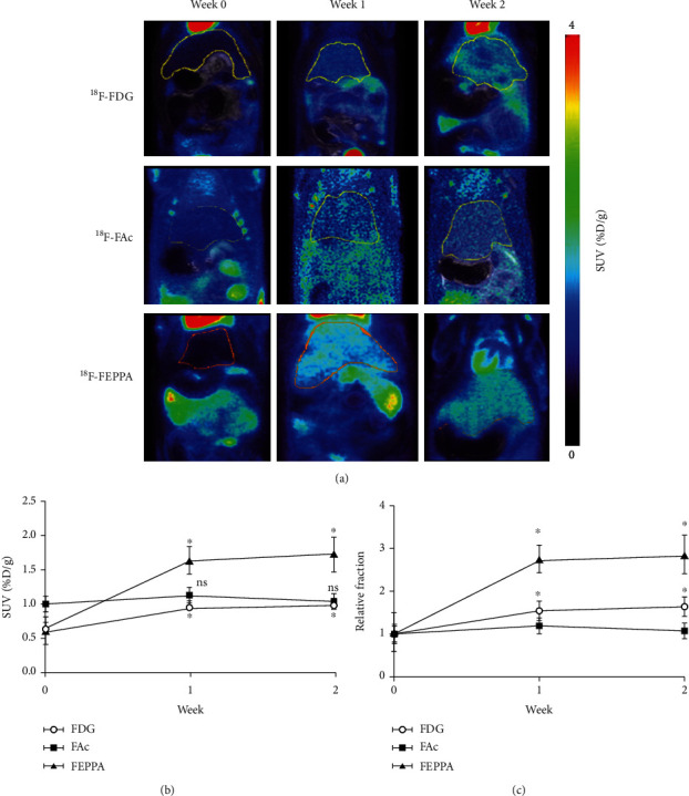
Developing sensitive diagnostic methods for a longitudinal evaluation of the status of liver fibrosis is a priority. This study is aimed at assessing the significance of longitudinal positron emission tomography (PET) imaging with 18F-labeling tracers for assessing liver fibrosis in a rat model with bile duct ligation (BDL). Twenty-one 6-week-old Sprague-Dawley male rats were used in this study. Longitudinal PET images using [18F]N-2-(2-fluoroethoxy)benzyl)-N-(4-phenoxypyridin-3-yl)acetamide ([18F]FEPPA) (n = 3), [18F]fluoroacetate ([18F]FAc) (n = 3), and 18F-fluoro-2-deoxy-D-glucose ([18F]FDG) (n = 3) were obtained at 0, 1, and 2 weeks after BDL. Biochemical assays, histological assays, immunohistochemical staining assays, and next generation sequencing analyses were also performed at 0 (n = 3), 1 (n = 3), 2 (n = 3), and 3 (n = 3) weeks after BDL, which demonstrated the severe damage in rat livers after BDL. Regarding [18F]FEPPA and [18F]FDG, there was a significantly higher uptake in the liver after BDL (both P < 0.05), which lasted until week 2. However, the uptake of [18F]FAc in the liver was not significantly different before and after BDL (P = 0.28). Collectively, both [18F]FEPPA and [18F]FDG can serve as sensitive probes for detecting the liver fibrosis. However, [18F]FAc is not recommended to diagnose liver fibrosis.
Molecular ImagingBiochemistry, Genetics and Molecular Biology-Biotechnology
自引率
3.60%
发文量
21
期刊介绍:
Molecular Imaging is a peer-reviewed, open access journal highlighting the breadth of molecular imaging research from basic science to preclinical studies to human applications. This serves both the scientific and clinical communities by disseminating novel results and concepts relevant to the biological study of normal and disease processes in both basic and translational studies ranging from mice to humans.
 求助内容:
求助内容: 应助结果提醒方式:


