{"title":"支架置入术后原发性主动脉肉瘤合并肿瘤梗死1例。","authors":"Hiroki Nakamura, Akihiko Kanki, Hiroyuki Watanabe, Kentarou Ono, Noriaki Kuwada, Shinsuke Saisho, Hirotake Nishimura, Akira Yamamoto, Tsutomu Tamada","doi":"10.1177/20584601211063360","DOIUrl":null,"url":null,"abstract":"<p><p>Primary aortic sarcoma is a very rare disease, and most primary aortic tumors are malignant mesenchymal tumors. We present the case of a 62-year-old man with sudden epigastric and back pain. Contrast-enhanced computed tomography (CT) revealed a mass lesion about 33.8 mm in diameter, in contact with the left side of the abdominal aorta. Impending rupture of an abdominal aortic aneurysm was suspected, so cardiovascular surgery for stent graft placement was performed the same day. Symptoms immediately improved and CT at 3 months postoperatively showed a marked decrease in lesion size, but the lesion subsequently grew again. Fluorodeoxyglucose (FDG)-positron emission tomography/CT was performed due to the possibility of malignant solid tumor, revealing markedly increased FDG accumulation (maximum standardized uptake value, 36.95) in the mass lesion. Primary aortic sarcoma was diagnosed from thoracoscopic biopsy. Here, we report a primary aortic sarcoma that shrank due to tumor infarction after stent graft placement, followed by tumor regrowth.</p>","PeriodicalId":72063,"journal":{"name":"Acta radiologica open","volume":"10 11","pages":"20584601211063360"},"PeriodicalIF":0.9000,"publicationDate":"2021-12-07","publicationTypes":"Journal Article","fieldsOfStudy":null,"isOpenAccess":false,"openAccessPdf":"https://ftp.ncbi.nlm.nih.gov/pub/pmc/oa_pdf/ea/0e/10.1177_20584601211063360.PMC8664319.pdf","citationCount":"0","resultStr":"{\"title\":\"A case of primary aortic sarcoma with tumor infarction after stent graft placement.\",\"authors\":\"Hiroki Nakamura, Akihiko Kanki, Hiroyuki Watanabe, Kentarou Ono, Noriaki Kuwada, Shinsuke Saisho, Hirotake Nishimura, Akira Yamamoto, Tsutomu Tamada\",\"doi\":\"10.1177/20584601211063360\",\"DOIUrl\":null,\"url\":null,\"abstract\":\"<p><p>Primary aortic sarcoma is a very rare disease, and most primary aortic tumors are malignant mesenchymal tumors. We present the case of a 62-year-old man with sudden epigastric and back pain. Contrast-enhanced computed tomography (CT) revealed a mass lesion about 33.8 mm in diameter, in contact with the left side of the abdominal aorta. Impending rupture of an abdominal aortic aneurysm was suspected, so cardiovascular surgery for stent graft placement was performed the same day. Symptoms immediately improved and CT at 3 months postoperatively showed a marked decrease in lesion size, but the lesion subsequently grew again. Fluorodeoxyglucose (FDG)-positron emission tomography/CT was performed due to the possibility of malignant solid tumor, revealing markedly increased FDG accumulation (maximum standardized uptake value, 36.95) in the mass lesion. Primary aortic sarcoma was diagnosed from thoracoscopic biopsy. Here, we report a primary aortic sarcoma that shrank due to tumor infarction after stent graft placement, followed by tumor regrowth.</p>\",\"PeriodicalId\":72063,\"journal\":{\"name\":\"Acta radiologica open\",\"volume\":\"10 11\",\"pages\":\"20584601211063360\"},\"PeriodicalIF\":0.9000,\"publicationDate\":\"2021-12-07\",\"publicationTypes\":\"Journal Article\",\"fieldsOfStudy\":null,\"isOpenAccess\":false,\"openAccessPdf\":\"https://ftp.ncbi.nlm.nih.gov/pub/pmc/oa_pdf/ea/0e/10.1177_20584601211063360.PMC8664319.pdf\",\"citationCount\":\"0\",\"resultStr\":null,\"platform\":\"Semanticscholar\",\"paperid\":null,\"PeriodicalName\":\"Acta radiologica open\",\"FirstCategoryId\":\"1085\",\"ListUrlMain\":\"https://doi.org/10.1177/20584601211063360\",\"RegionNum\":0,\"RegionCategory\":null,\"ArticlePicture\":[],\"TitleCN\":null,\"AbstractTextCN\":null,\"PMCID\":null,\"EPubDate\":\"2021/11/1 0:00:00\",\"PubModel\":\"eCollection\",\"JCR\":\"Q4\",\"JCRName\":\"RADIOLOGY, NUCLEAR MEDICINE & MEDICAL IMAGING\",\"Score\":null,\"Total\":0}","platform":"Semanticscholar","paperid":null,"PeriodicalName":"Acta radiologica open","FirstCategoryId":"1085","ListUrlMain":"https://doi.org/10.1177/20584601211063360","RegionNum":0,"RegionCategory":null,"ArticlePicture":[],"TitleCN":null,"AbstractTextCN":null,"PMCID":null,"EPubDate":"2021/11/1 0:00:00","PubModel":"eCollection","JCR":"Q4","JCRName":"RADIOLOGY, NUCLEAR MEDICINE & MEDICAL IMAGING","Score":null,"Total":0}
A case of primary aortic sarcoma with tumor infarction after stent graft placement.
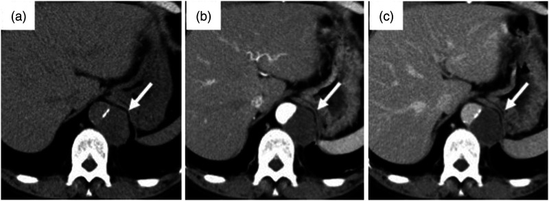
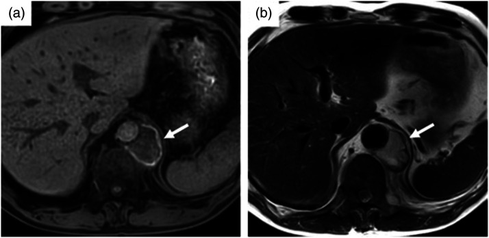
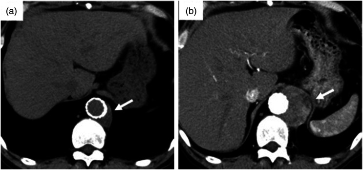
Primary aortic sarcoma is a very rare disease, and most primary aortic tumors are malignant mesenchymal tumors. We present the case of a 62-year-old man with sudden epigastric and back pain. Contrast-enhanced computed tomography (CT) revealed a mass lesion about 33.8 mm in diameter, in contact with the left side of the abdominal aorta. Impending rupture of an abdominal aortic aneurysm was suspected, so cardiovascular surgery for stent graft placement was performed the same day. Symptoms immediately improved and CT at 3 months postoperatively showed a marked decrease in lesion size, but the lesion subsequently grew again. Fluorodeoxyglucose (FDG)-positron emission tomography/CT was performed due to the possibility of malignant solid tumor, revealing markedly increased FDG accumulation (maximum standardized uptake value, 36.95) in the mass lesion. Primary aortic sarcoma was diagnosed from thoracoscopic biopsy. Here, we report a primary aortic sarcoma that shrank due to tumor infarction after stent graft placement, followed by tumor regrowth.

 求助内容:
求助内容: 应助结果提醒方式:


