Shelley S Mason, Sean S Kohles, Shelley R Winn, Randy D Zelick
{"title":"维生素 D3 在工程成骨细胞前体细胞系中的肝外 25-羟化,探索其对骨骼发育过程中细胞增殖和基质成熟的影响。","authors":"Shelley S Mason, Sean S Kohles, Shelley R Winn, Randy D Zelick","doi":"10.1155/2013/956362","DOIUrl":null,"url":null,"abstract":"<p><p>Osteoblastic precursors experience distinct stages during differentiation and bone development, which include proliferation, extracellular matrix (ECM) maturation, and ECM mineralization. It is well known that vitamin D plays a large role in the regulation of bone mineralization and homeostasis via the endocrine system. The activation of vitamin D requires two sequential hydroxylation steps, first in the kidney and then in the liver, in order to carry out its role in calcium homeostasis. Recent research has demonstrated that human-derived mesenchymal stem cells (MSCs) and osteoblasts can metabolize the immediate vitamin D precursor 25-dihydroxyvitamin D<sub>3</sub> (25OHD<sub>3</sub>) to the active steroid l<i>α</i>,25-dihydroxyvitamin D<sub>3</sub> (1,25OH<sub>2</sub>D<sub>3</sub>) and elicit an osteogenic response. However, reports of extrahepatic metabolism of vitamin D<sub>3</sub>, the parental vitamin D precursor, have been limited. In this study, we investigated whether osteoblast precursors have the capacity to convert vitamin D<sub>3</sub> to 1,25OH<sub>2</sub>D<sub>3</sub> and examined the potential of vitamin D<sub>3</sub> to induce 1,25OH<sub>2</sub>D<sub>3</sub> associated biological activities in osteoblast precursors. It was demonstrated that the engineered osteoblast precursor derived from human marrow (OPC1) is capable of metabolizing vitamin D<sub>3</sub> to 1,25OH<sub>2</sub>D<sub>3</sub> in a dose-dependent manner. It was also demonstrated that administration of vitamin D<sub>3</sub> leads to the increase in alkaline phosphatase (ALP) activity associated with osteoblast ECM maturation and calcium deposits and a decrease in cellular proliferation in both osteoblast precursor cell lines 0PC1 andOMC3T3-E1. These findings provide a two-dimensional culture foundation for future three-dimensional engineered tissue studies using the OPC1 cell line.</p>","PeriodicalId":93456,"journal":{"name":"ISRN biomedical engineering","volume":"2013 ","pages":""},"PeriodicalIF":0.0000,"publicationDate":"2013-01-01","publicationTypes":"Journal Article","fieldsOfStudy":null,"isOpenAccess":false,"openAccessPdf":"https://www.ncbi.nlm.nih.gov/pmc/articles/PMC8667671/pdf/","citationCount":"0","resultStr":"{\"title\":\"Extrahepatic 25-Hydroxylation of Vitamin D<sub>3</sub> in an Engineered Osteoblast Precursor Cell Line Exploring the Influence on Cellular Proliferation and Matrix Maturation during Bone Development.\",\"authors\":\"Shelley S Mason, Sean S Kohles, Shelley R Winn, Randy D Zelick\",\"doi\":\"10.1155/2013/956362\",\"DOIUrl\":null,\"url\":null,\"abstract\":\"<p><p>Osteoblastic precursors experience distinct stages during differentiation and bone development, which include proliferation, extracellular matrix (ECM) maturation, and ECM mineralization. It is well known that vitamin D plays a large role in the regulation of bone mineralization and homeostasis via the endocrine system. The activation of vitamin D requires two sequential hydroxylation steps, first in the kidney and then in the liver, in order to carry out its role in calcium homeostasis. Recent research has demonstrated that human-derived mesenchymal stem cells (MSCs) and osteoblasts can metabolize the immediate vitamin D precursor 25-dihydroxyvitamin D<sub>3</sub> (25OHD<sub>3</sub>) to the active steroid l<i>α</i>,25-dihydroxyvitamin D<sub>3</sub> (1,25OH<sub>2</sub>D<sub>3</sub>) and elicit an osteogenic response. However, reports of extrahepatic metabolism of vitamin D<sub>3</sub>, the parental vitamin D precursor, have been limited. In this study, we investigated whether osteoblast precursors have the capacity to convert vitamin D<sub>3</sub> to 1,25OH<sub>2</sub>D<sub>3</sub> and examined the potential of vitamin D<sub>3</sub> to induce 1,25OH<sub>2</sub>D<sub>3</sub> associated biological activities in osteoblast precursors. It was demonstrated that the engineered osteoblast precursor derived from human marrow (OPC1) is capable of metabolizing vitamin D<sub>3</sub> to 1,25OH<sub>2</sub>D<sub>3</sub> in a dose-dependent manner. It was also demonstrated that administration of vitamin D<sub>3</sub> leads to the increase in alkaline phosphatase (ALP) activity associated with osteoblast ECM maturation and calcium deposits and a decrease in cellular proliferation in both osteoblast precursor cell lines 0PC1 andOMC3T3-E1. These findings provide a two-dimensional culture foundation for future three-dimensional engineered tissue studies using the OPC1 cell line.</p>\",\"PeriodicalId\":93456,\"journal\":{\"name\":\"ISRN biomedical engineering\",\"volume\":\"2013 \",\"pages\":\"\"},\"PeriodicalIF\":0.0000,\"publicationDate\":\"2013-01-01\",\"publicationTypes\":\"Journal Article\",\"fieldsOfStudy\":null,\"isOpenAccess\":false,\"openAccessPdf\":\"https://www.ncbi.nlm.nih.gov/pmc/articles/PMC8667671/pdf/\",\"citationCount\":\"0\",\"resultStr\":null,\"platform\":\"Semanticscholar\",\"paperid\":null,\"PeriodicalName\":\"ISRN biomedical engineering\",\"FirstCategoryId\":\"1085\",\"ListUrlMain\":\"https://doi.org/10.1155/2013/956362\",\"RegionNum\":0,\"RegionCategory\":null,\"ArticlePicture\":[],\"TitleCN\":null,\"AbstractTextCN\":null,\"PMCID\":null,\"EPubDate\":\"2013/6/4 0:00:00\",\"PubModel\":\"Epub\",\"JCR\":\"\",\"JCRName\":\"\",\"Score\":null,\"Total\":0}","platform":"Semanticscholar","paperid":null,"PeriodicalName":"ISRN biomedical engineering","FirstCategoryId":"1085","ListUrlMain":"https://doi.org/10.1155/2013/956362","RegionNum":0,"RegionCategory":null,"ArticlePicture":[],"TitleCN":null,"AbstractTextCN":null,"PMCID":null,"EPubDate":"2013/6/4 0:00:00","PubModel":"Epub","JCR":"","JCRName":"","Score":null,"Total":0}
引用次数: 0
摘要
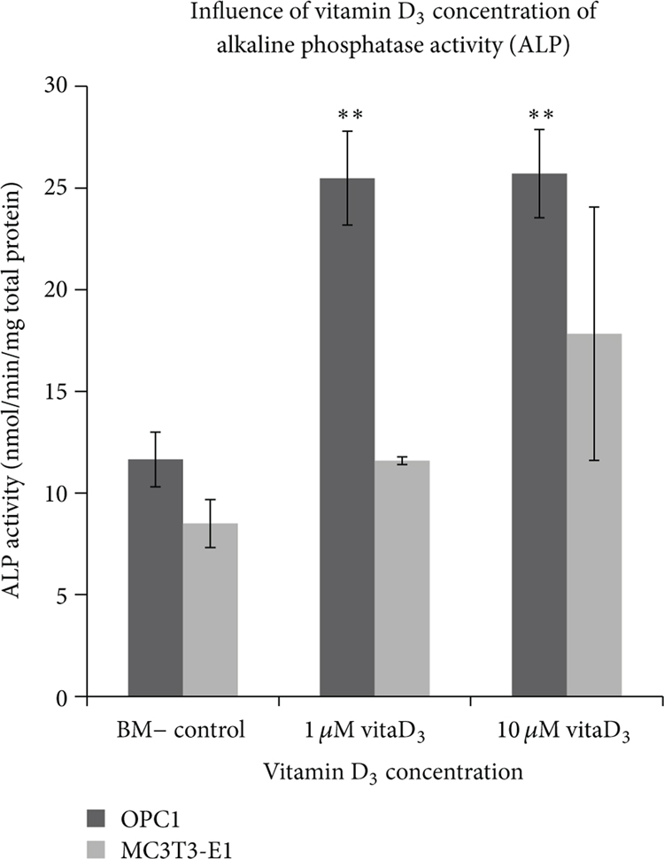
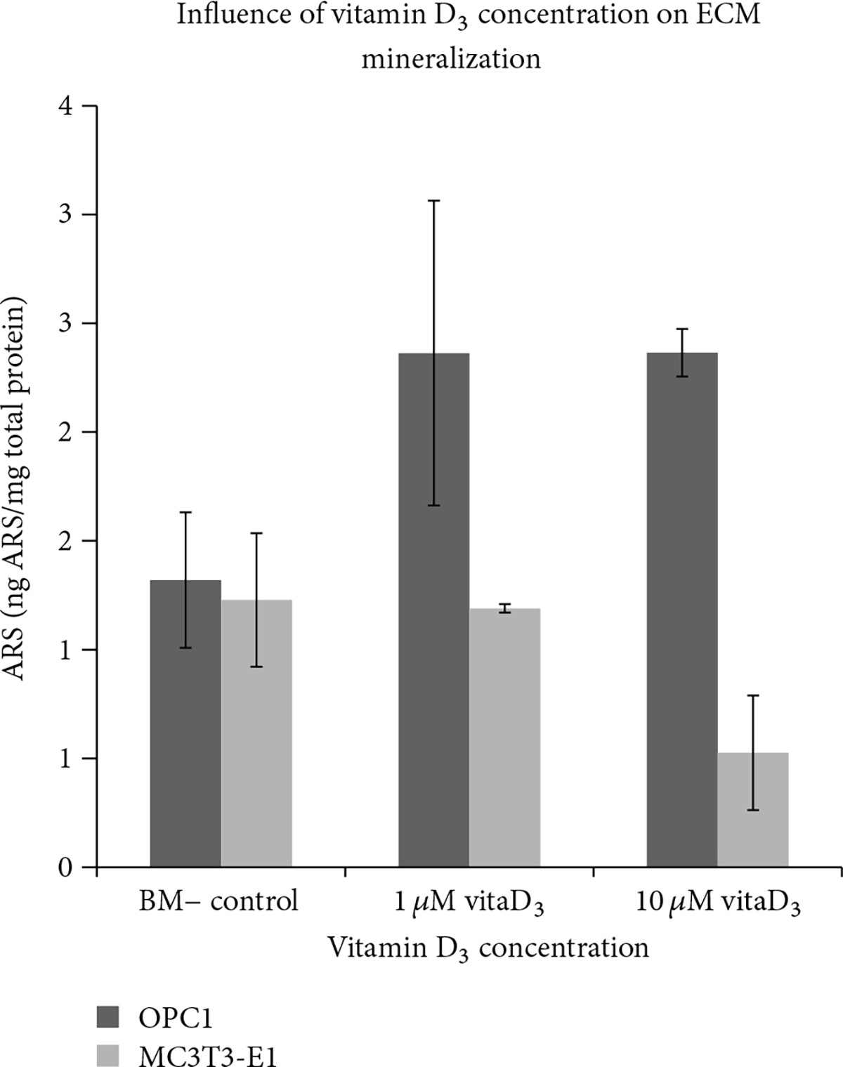
成骨细胞前体在分化和骨骼发育过程中经历了不同的阶段,包括增殖、细胞外基质(ECM)成熟和 ECM 矿化。众所周知,维生素 D 通过内分泌系统在调节骨矿化和平衡方面发挥着重要作用。维生素 D 的活化需要两个连续的羟化步骤,首先在肾脏,然后在肝脏,才能发挥其在钙平衡中的作用。最近的研究表明,人源间充质干细胞(MSCs)和成骨细胞可将维生素 D 的直接前体 25-二羟维生素 D3(25OHD3)代谢为活性类固醇 lα,25-二羟维生素 D3(1,25OH2D3)并引起成骨反应。然而,有关维生素 D3(母体维生素 D 的前体)肝外代谢的报道十分有限。在这项研究中,我们研究了成骨细胞前体是否有能力将维生素 D3 转化为 1,25OH2D3,并考察了维生素 D3 在成骨细胞前体中诱导 1,25OH2D3 相关生物活性的潜力。研究表明,源自人类骨髓的工程成骨细胞前体(OPC1)能够以剂量依赖的方式将维生素 D3 代谢为 1,25OH2D3。研究还证明,在成骨细胞前体细胞系 0PC1 和OMC3T3-E1 中,服用维生素 D3 会导致与成骨细胞 ECM 成熟和钙沉积有关的碱性磷酸酶(ALP)活性增加,并导致细胞增殖减少。这些发现为将来使用 OPC1 细胞系进行三维工程组织研究奠定了二维培养基础。
Extrahepatic 25-Hydroxylation of Vitamin D3 in an Engineered Osteoblast Precursor Cell Line Exploring the Influence on Cellular Proliferation and Matrix Maturation during Bone Development.
Osteoblastic precursors experience distinct stages during differentiation and bone development, which include proliferation, extracellular matrix (ECM) maturation, and ECM mineralization. It is well known that vitamin D plays a large role in the regulation of bone mineralization and homeostasis via the endocrine system. The activation of vitamin D requires two sequential hydroxylation steps, first in the kidney and then in the liver, in order to carry out its role in calcium homeostasis. Recent research has demonstrated that human-derived mesenchymal stem cells (MSCs) and osteoblasts can metabolize the immediate vitamin D precursor 25-dihydroxyvitamin D3 (25OHD3) to the active steroid lα,25-dihydroxyvitamin D3 (1,25OH2D3) and elicit an osteogenic response. However, reports of extrahepatic metabolism of vitamin D3, the parental vitamin D precursor, have been limited. In this study, we investigated whether osteoblast precursors have the capacity to convert vitamin D3 to 1,25OH2D3 and examined the potential of vitamin D3 to induce 1,25OH2D3 associated biological activities in osteoblast precursors. It was demonstrated that the engineered osteoblast precursor derived from human marrow (OPC1) is capable of metabolizing vitamin D3 to 1,25OH2D3 in a dose-dependent manner. It was also demonstrated that administration of vitamin D3 leads to the increase in alkaline phosphatase (ALP) activity associated with osteoblast ECM maturation and calcium deposits and a decrease in cellular proliferation in both osteoblast precursor cell lines 0PC1 andOMC3T3-E1. These findings provide a two-dimensional culture foundation for future three-dimensional engineered tissue studies using the OPC1 cell line.


 求助内容:
求助内容: 应助结果提醒方式:


