Rieke L Meister, Michael Groth, Julian H W Jürgens, Shuo Zhang, Jan H Buhk, Jochen Herrmann
{"title":"压缩感在儿童脑肿瘤磁共振成像中的应用:图像质量、检查时间和能量释放的评估。","authors":"Rieke L Meister, Michael Groth, Julian H W Jürgens, Shuo Zhang, Jan H Buhk, Jochen Herrmann","doi":"10.1007/s00062-021-01112-3","DOIUrl":null,"url":null,"abstract":"<p><strong>Purpose: </strong>To compare the image quality, examination time, and total energy release of a standardized pediatric brain tumor magnetic resonance imaging (MRI) protocol performed with and without compressed sensitivity encoding (C-SENSE). Recently introduced as an acceleration technique in MRI, we hypothesized that C‑SENSE would improve image quality, reduce the examination time and radiofrequency-induced energy release compared with conventional examination in a pediatric brain tumor protocol.</p><p><strong>Methods: </strong>This retrospective study included 22 patients aged 2.33-18.83 years with different brain tumor types who had previously undergone conventional MRI examination and underwent follow-up C‑SENSE examination. Both examinations were conducted with a 3.0-Tesla device and included pre-contrast and post-contrast T1-weighted turbo-field-echo, T2-weighted turbo-spin-echo, and fluid-attenuated inversion recovery sequences. Image quality was assessed in four anatomical regions of interest (tumor area, cerebral cortex, basal ganglia, and posterior fossa) using a 5-point scale. Reader preference between the standard and C‑SENSE images was evaluated. The total examination duration and energy deposit were compared based on scanner log file analysis.</p><p><strong>Results: </strong>Relative to standard examinations, C‑SENSE examinations were characterized by shorter total examination times (26.1 ± 3.93 vs. 22.18 ± 2.31 min; P = 0.001), reduced total energy deposit (206.0 ± 19.7 vs. 92.3 ± 18.2 J/kg; P < 0.001), and higher image quality (overall P < 0.001).</p><p><strong>Conclusion: </strong>C‑SENSE contributes to the improvement of image quality, reduction of scan times and radiofrequency-induced energy release relative to the standard protocol in pediatric brain tumor MRI.</p>","PeriodicalId":49298,"journal":{"name":"Clinical Neuroradiology","volume":"32 3","pages":"725-733"},"PeriodicalIF":2.4000,"publicationDate":"2022-09-01","publicationTypes":"Journal Article","fieldsOfStudy":null,"isOpenAccess":false,"openAccessPdf":"https://www.ncbi.nlm.nih.gov/pmc/articles/PMC9424145/pdf/","citationCount":"8","resultStr":"{\"title\":\"Compressed SENSE in Pediatric Brain Tumor MR Imaging : Assessment of Image Quality, Examination Time and Energy Release.\",\"authors\":\"Rieke L Meister, Michael Groth, Julian H W Jürgens, Shuo Zhang, Jan H Buhk, Jochen Herrmann\",\"doi\":\"10.1007/s00062-021-01112-3\",\"DOIUrl\":null,\"url\":null,\"abstract\":\"<p><strong>Purpose: </strong>To compare the image quality, examination time, and total energy release of a standardized pediatric brain tumor magnetic resonance imaging (MRI) protocol performed with and without compressed sensitivity encoding (C-SENSE). Recently introduced as an acceleration technique in MRI, we hypothesized that C‑SENSE would improve image quality, reduce the examination time and radiofrequency-induced energy release compared with conventional examination in a pediatric brain tumor protocol.</p><p><strong>Methods: </strong>This retrospective study included 22 patients aged 2.33-18.83 years with different brain tumor types who had previously undergone conventional MRI examination and underwent follow-up C‑SENSE examination. Both examinations were conducted with a 3.0-Tesla device and included pre-contrast and post-contrast T1-weighted turbo-field-echo, T2-weighted turbo-spin-echo, and fluid-attenuated inversion recovery sequences. Image quality was assessed in four anatomical regions of interest (tumor area, cerebral cortex, basal ganglia, and posterior fossa) using a 5-point scale. Reader preference between the standard and C‑SENSE images was evaluated. The total examination duration and energy deposit were compared based on scanner log file analysis.</p><p><strong>Results: </strong>Relative to standard examinations, C‑SENSE examinations were characterized by shorter total examination times (26.1 ± 3.93 vs. 22.18 ± 2.31 min; P = 0.001), reduced total energy deposit (206.0 ± 19.7 vs. 92.3 ± 18.2 J/kg; P < 0.001), and higher image quality (overall P < 0.001).</p><p><strong>Conclusion: </strong>C‑SENSE contributes to the improvement of image quality, reduction of scan times and radiofrequency-induced energy release relative to the standard protocol in pediatric brain tumor MRI.</p>\",\"PeriodicalId\":49298,\"journal\":{\"name\":\"Clinical Neuroradiology\",\"volume\":\"32 3\",\"pages\":\"725-733\"},\"PeriodicalIF\":2.4000,\"publicationDate\":\"2022-09-01\",\"publicationTypes\":\"Journal Article\",\"fieldsOfStudy\":null,\"isOpenAccess\":false,\"openAccessPdf\":\"https://www.ncbi.nlm.nih.gov/pmc/articles/PMC9424145/pdf/\",\"citationCount\":\"8\",\"resultStr\":null,\"platform\":\"Semanticscholar\",\"paperid\":null,\"PeriodicalName\":\"Clinical Neuroradiology\",\"FirstCategoryId\":\"3\",\"ListUrlMain\":\"https://doi.org/10.1007/s00062-021-01112-3\",\"RegionNum\":3,\"RegionCategory\":\"医学\",\"ArticlePicture\":[],\"TitleCN\":null,\"AbstractTextCN\":null,\"PMCID\":null,\"EPubDate\":\"2022/1/7 0:00:00\",\"PubModel\":\"Epub\",\"JCR\":\"Q2\",\"JCRName\":\"CLINICAL NEUROLOGY\",\"Score\":null,\"Total\":0}","platform":"Semanticscholar","paperid":null,"PeriodicalName":"Clinical Neuroradiology","FirstCategoryId":"3","ListUrlMain":"https://doi.org/10.1007/s00062-021-01112-3","RegionNum":3,"RegionCategory":"医学","ArticlePicture":[],"TitleCN":null,"AbstractTextCN":null,"PMCID":null,"EPubDate":"2022/1/7 0:00:00","PubModel":"Epub","JCR":"Q2","JCRName":"CLINICAL NEUROLOGY","Score":null,"Total":0}
Compressed SENSE in Pediatric Brain Tumor MR Imaging : Assessment of Image Quality, Examination Time and Energy Release.
Purpose: To compare the image quality, examination time, and total energy release of a standardized pediatric brain tumor magnetic resonance imaging (MRI) protocol performed with and without compressed sensitivity encoding (C-SENSE). Recently introduced as an acceleration technique in MRI, we hypothesized that C‑SENSE would improve image quality, reduce the examination time and radiofrequency-induced energy release compared with conventional examination in a pediatric brain tumor protocol.
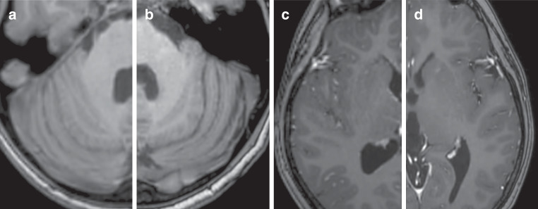
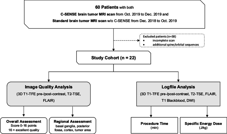
Methods: This retrospective study included 22 patients aged 2.33-18.83 years with different brain tumor types who had previously undergone conventional MRI examination and underwent follow-up C‑SENSE examination. Both examinations were conducted with a 3.0-Tesla device and included pre-contrast and post-contrast T1-weighted turbo-field-echo, T2-weighted turbo-spin-echo, and fluid-attenuated inversion recovery sequences. Image quality was assessed in four anatomical regions of interest (tumor area, cerebral cortex, basal ganglia, and posterior fossa) using a 5-point scale. Reader preference between the standard and C‑SENSE images was evaluated. The total examination duration and energy deposit were compared based on scanner log file analysis.
Results: Relative to standard examinations, C‑SENSE examinations were characterized by shorter total examination times (26.1 ± 3.93 vs. 22.18 ± 2.31 min; P = 0.001), reduced total energy deposit (206.0 ± 19.7 vs. 92.3 ± 18.2 J/kg; P < 0.001), and higher image quality (overall P < 0.001).
Conclusion: C‑SENSE contributes to the improvement of image quality, reduction of scan times and radiofrequency-induced energy release relative to the standard protocol in pediatric brain tumor MRI.
期刊介绍:
Clinical Neuroradiology provides current information, original contributions, and reviews in the field of neuroradiology. An interdisciplinary approach is accomplished by diagnostic and therapeutic contributions related to associated subjects.
The international coverage and relevance of the journal is underlined by its being the official journal of the German, Swiss, and Austrian Societies of Neuroradiology.

 求助内容:
求助内容: 应助结果提醒方式:


