A I Korsakova, I A Zhadobova, A S Klochkov, S A Durnovo, A V Kochubeynik, E A Durnovo
{"title":"改良两段式劈开技术在水平萎缩后颌骨控制嵴增大中的应用研究。","authors":"A I Korsakova, I A Zhadobova, A S Klochkov, S A Durnovo, A V Kochubeynik, E A Durnovo","doi":"10.17691/stm2020.12.4.05","DOIUrl":null,"url":null,"abstract":"<p><p>Various bone grafting methods are applied to eliminate horizontal atrophy of the jaws. However, problem complexity brings about ongoing research and development of new ways to achieve the predicted stable and long-term results of implantological treatment. <b>The aim of the study</b> was to evaluate the results of the developed method for bone grafting, a modified two-stage split technique for controlled ridge augmentation in horizontally atrophic posterior mandible, using radiological analysis data.</p><p><strong>Materials and methods: </strong>The study group included 18 patients with horizontally atrophic posterior mandible. According to cone beam computed tomography, 39 jaw segments were assessed before plastic surgery and after applying the two-stage split-crest technique for controlled ridge augmentation. The alveolar ridge width was estimated in the area of its top and at a distance of 1, 3, 5 mm from it using the vector of future implant position and taking into account the angle of inclination of the atrophic region of the mandible.</p><p><strong>Results: </strong>When analyzing edentulous areas in the posterior mandible before treatment, there was rather a large angle of lingual inclination of the alveolar ridge. After 6 months, the average increase in bone tissue width in the region of the alveolar ridge top was 82%, it was 50.6% at a height of 1 mm from the top of the crest, 58.8% at 3 mm height, 46.7% at 5 mm (p≤0.05). Certain patterns of bone tissue growth were revealed depending on the structure of the reconstructed area. The most significant results were obtained in the molar segments of the mandible.</p><p><strong>Conclusion: </strong>The developed modified two-stage split technique for alveolar ridge augmentation allows achieving the required volume of bone tissue in the posterior mandible for successful implant treatment.</p>","PeriodicalId":51886,"journal":{"name":"Sovremennye Tehnologii v Medicine","volume":"12 4","pages":"40-46"},"PeriodicalIF":0.9000,"publicationDate":"2021-01-01","publicationTypes":"Journal Article","fieldsOfStudy":null,"isOpenAccess":false,"openAccessPdf":"https://www.ncbi.nlm.nih.gov/pmc/articles/PMC8596280/pdf/","citationCount":"4","resultStr":"{\"title\":\"Modified Two-Stage Split Technique for Controlled Ridge Augmentation in Horizontally Atrophic Posterior Mandible: the First Stage of Research.\",\"authors\":\"A I Korsakova, I A Zhadobova, A S Klochkov, S A Durnovo, A V Kochubeynik, E A Durnovo\",\"doi\":\"10.17691/stm2020.12.4.05\",\"DOIUrl\":null,\"url\":null,\"abstract\":\"<p><p>Various bone grafting methods are applied to eliminate horizontal atrophy of the jaws. However, problem complexity brings about ongoing research and development of new ways to achieve the predicted stable and long-term results of implantological treatment. <b>The aim of the study</b> was to evaluate the results of the developed method for bone grafting, a modified two-stage split technique for controlled ridge augmentation in horizontally atrophic posterior mandible, using radiological analysis data.</p><p><strong>Materials and methods: </strong>The study group included 18 patients with horizontally atrophic posterior mandible. According to cone beam computed tomography, 39 jaw segments were assessed before plastic surgery and after applying the two-stage split-crest technique for controlled ridge augmentation. The alveolar ridge width was estimated in the area of its top and at a distance of 1, 3, 5 mm from it using the vector of future implant position and taking into account the angle of inclination of the atrophic region of the mandible.</p><p><strong>Results: </strong>When analyzing edentulous areas in the posterior mandible before treatment, there was rather a large angle of lingual inclination of the alveolar ridge. After 6 months, the average increase in bone tissue width in the region of the alveolar ridge top was 82%, it was 50.6% at a height of 1 mm from the top of the crest, 58.8% at 3 mm height, 46.7% at 5 mm (p≤0.05). Certain patterns of bone tissue growth were revealed depending on the structure of the reconstructed area. The most significant results were obtained in the molar segments of the mandible.</p><p><strong>Conclusion: </strong>The developed modified two-stage split technique for alveolar ridge augmentation allows achieving the required volume of bone tissue in the posterior mandible for successful implant treatment.</p>\",\"PeriodicalId\":51886,\"journal\":{\"name\":\"Sovremennye Tehnologii v Medicine\",\"volume\":\"12 4\",\"pages\":\"40-46\"},\"PeriodicalIF\":0.9000,\"publicationDate\":\"2021-01-01\",\"publicationTypes\":\"Journal Article\",\"fieldsOfStudy\":null,\"isOpenAccess\":false,\"openAccessPdf\":\"https://www.ncbi.nlm.nih.gov/pmc/articles/PMC8596280/pdf/\",\"citationCount\":\"4\",\"resultStr\":null,\"platform\":\"Semanticscholar\",\"paperid\":null,\"PeriodicalName\":\"Sovremennye Tehnologii v Medicine\",\"FirstCategoryId\":\"1085\",\"ListUrlMain\":\"https://doi.org/10.17691/stm2020.12.4.05\",\"RegionNum\":0,\"RegionCategory\":null,\"ArticlePicture\":[],\"TitleCN\":null,\"AbstractTextCN\":null,\"PMCID\":null,\"EPubDate\":\"2020/8/27 0:00:00\",\"PubModel\":\"Epub\",\"JCR\":\"Q4\",\"JCRName\":\"MEDICINE, RESEARCH & EXPERIMENTAL\",\"Score\":null,\"Total\":0}","platform":"Semanticscholar","paperid":null,"PeriodicalName":"Sovremennye Tehnologii v Medicine","FirstCategoryId":"1085","ListUrlMain":"https://doi.org/10.17691/stm2020.12.4.05","RegionNum":0,"RegionCategory":null,"ArticlePicture":[],"TitleCN":null,"AbstractTextCN":null,"PMCID":null,"EPubDate":"2020/8/27 0:00:00","PubModel":"Epub","JCR":"Q4","JCRName":"MEDICINE, RESEARCH & EXPERIMENTAL","Score":null,"Total":0}
Modified Two-Stage Split Technique for Controlled Ridge Augmentation in Horizontally Atrophic Posterior Mandible: the First Stage of Research.
Various bone grafting methods are applied to eliminate horizontal atrophy of the jaws. However, problem complexity brings about ongoing research and development of new ways to achieve the predicted stable and long-term results of implantological treatment. The aim of the study was to evaluate the results of the developed method for bone grafting, a modified two-stage split technique for controlled ridge augmentation in horizontally atrophic posterior mandible, using radiological analysis data.
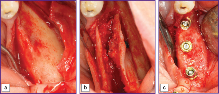
Materials and methods: The study group included 18 patients with horizontally atrophic posterior mandible. According to cone beam computed tomography, 39 jaw segments were assessed before plastic surgery and after applying the two-stage split-crest technique for controlled ridge augmentation. The alveolar ridge width was estimated in the area of its top and at a distance of 1, 3, 5 mm from it using the vector of future implant position and taking into account the angle of inclination of the atrophic region of the mandible.
Results: When analyzing edentulous areas in the posterior mandible before treatment, there was rather a large angle of lingual inclination of the alveolar ridge. After 6 months, the average increase in bone tissue width in the region of the alveolar ridge top was 82%, it was 50.6% at a height of 1 mm from the top of the crest, 58.8% at 3 mm height, 46.7% at 5 mm (p≤0.05). Certain patterns of bone tissue growth were revealed depending on the structure of the reconstructed area. The most significant results were obtained in the molar segments of the mandible.
Conclusion: The developed modified two-stage split technique for alveolar ridge augmentation allows achieving the required volume of bone tissue in the posterior mandible for successful implant treatment.


 求助内容:
求助内容: 应助结果提醒方式:


