Oscar Otero-Marquez, Mona Fayad, Alexander Pinhas, Toco Y P Chui, Richard B Rosen, Harsha S Reddy
{"title":"Teprotumumab治疗前后甲状腺眼病视网膜表面巨噬细胞的变化。","authors":"Oscar Otero-Marquez, Mona Fayad, Alexander Pinhas, Toco Y P Chui, Richard B Rosen, Harsha S Reddy","doi":"10.1155/2022/5275309","DOIUrl":null,"url":null,"abstract":"<p><p>Retinal surface macrophages play key roles in the regulation of immune response, maintenance of vitreous clarity, and tissue repair. We examined the variation of parafoveal surface macrophages in a thyroid eye disease (TED) patient before and after treatment with teprotumumab (Tepezza, Horizon therapeutics). Pre- and posttreatment parafoveal surface macrophages were imaged using clinical <i>en face</i> OCT, and their density was assessed using a novel cell density mapping technique. Pretreatment, surface macrophage cell density was high. Macrophages had a nonuniform spatial distribution, and their appearance was round with few protrusions, consistent with an \"activated\" state. Posttreatment, cell density decreased. The macrophages were regularly spaced and had a ramified appearance and filopodia-like processes, consistent with a \"quiescent\" state. Surface macrophage density decreased as the Clinical Activity Score (CAS) decreased with teprotumumab treatment, suggesting a potential association of these cells with an underlying intraocular and retinal inflammatory process previously not described in TED.</p>","PeriodicalId":9603,"journal":{"name":"Case Reports in Ophthalmological Medicine","volume":null,"pages":null},"PeriodicalIF":0.7000,"publicationDate":"2022-02-07","publicationTypes":"Journal Article","fieldsOfStudy":null,"isOpenAccess":false,"openAccessPdf":"https://www.ncbi.nlm.nih.gov/pmc/articles/PMC8844343/pdf/","citationCount":"2","resultStr":"{\"title\":\"Retinal Surface Macrophage Changes in Thyroid Eye Disease before and after Treatment with Teprotumumab.\",\"authors\":\"Oscar Otero-Marquez, Mona Fayad, Alexander Pinhas, Toco Y P Chui, Richard B Rosen, Harsha S Reddy\",\"doi\":\"10.1155/2022/5275309\",\"DOIUrl\":null,\"url\":null,\"abstract\":\"<p><p>Retinal surface macrophages play key roles in the regulation of immune response, maintenance of vitreous clarity, and tissue repair. We examined the variation of parafoveal surface macrophages in a thyroid eye disease (TED) patient before and after treatment with teprotumumab (Tepezza, Horizon therapeutics). Pre- and posttreatment parafoveal surface macrophages were imaged using clinical <i>en face</i> OCT, and their density was assessed using a novel cell density mapping technique. Pretreatment, surface macrophage cell density was high. Macrophages had a nonuniform spatial distribution, and their appearance was round with few protrusions, consistent with an \\\"activated\\\" state. Posttreatment, cell density decreased. The macrophages were regularly spaced and had a ramified appearance and filopodia-like processes, consistent with a \\\"quiescent\\\" state. Surface macrophage density decreased as the Clinical Activity Score (CAS) decreased with teprotumumab treatment, suggesting a potential association of these cells with an underlying intraocular and retinal inflammatory process previously not described in TED.</p>\",\"PeriodicalId\":9603,\"journal\":{\"name\":\"Case Reports in Ophthalmological Medicine\",\"volume\":null,\"pages\":null},\"PeriodicalIF\":0.7000,\"publicationDate\":\"2022-02-07\",\"publicationTypes\":\"Journal Article\",\"fieldsOfStudy\":null,\"isOpenAccess\":false,\"openAccessPdf\":\"https://www.ncbi.nlm.nih.gov/pmc/articles/PMC8844343/pdf/\",\"citationCount\":\"2\",\"resultStr\":null,\"platform\":\"Semanticscholar\",\"paperid\":null,\"PeriodicalName\":\"Case Reports in Ophthalmological Medicine\",\"FirstCategoryId\":\"1085\",\"ListUrlMain\":\"https://doi.org/10.1155/2022/5275309\",\"RegionNum\":0,\"RegionCategory\":null,\"ArticlePicture\":[],\"TitleCN\":null,\"AbstractTextCN\":null,\"PMCID\":null,\"EPubDate\":\"2022/1/1 0:00:00\",\"PubModel\":\"eCollection\",\"JCR\":\"Q4\",\"JCRName\":\"OPHTHALMOLOGY\",\"Score\":null,\"Total\":0}","platform":"Semanticscholar","paperid":null,"PeriodicalName":"Case Reports in Ophthalmological Medicine","FirstCategoryId":"1085","ListUrlMain":"https://doi.org/10.1155/2022/5275309","RegionNum":0,"RegionCategory":null,"ArticlePicture":[],"TitleCN":null,"AbstractTextCN":null,"PMCID":null,"EPubDate":"2022/1/1 0:00:00","PubModel":"eCollection","JCR":"Q4","JCRName":"OPHTHALMOLOGY","Score":null,"Total":0}
Retinal Surface Macrophage Changes in Thyroid Eye Disease before and after Treatment with Teprotumumab.
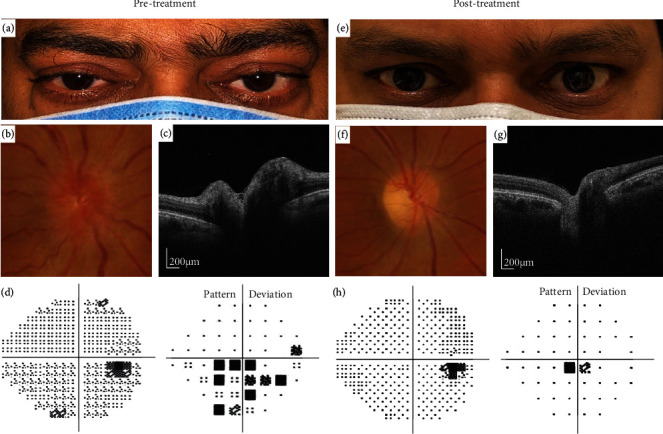
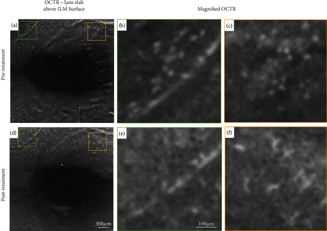
Retinal surface macrophages play key roles in the regulation of immune response, maintenance of vitreous clarity, and tissue repair. We examined the variation of parafoveal surface macrophages in a thyroid eye disease (TED) patient before and after treatment with teprotumumab (Tepezza, Horizon therapeutics). Pre- and posttreatment parafoveal surface macrophages were imaged using clinical en face OCT, and their density was assessed using a novel cell density mapping technique. Pretreatment, surface macrophage cell density was high. Macrophages had a nonuniform spatial distribution, and their appearance was round with few protrusions, consistent with an "activated" state. Posttreatment, cell density decreased. The macrophages were regularly spaced and had a ramified appearance and filopodia-like processes, consistent with a "quiescent" state. Surface macrophage density decreased as the Clinical Activity Score (CAS) decreased with teprotumumab treatment, suggesting a potential association of these cells with an underlying intraocular and retinal inflammatory process previously not described in TED.


 求助内容:
求助内容: 应助结果提醒方式:


