Bhargavi Pawar, Suneetha N Lobo, Mary Joseph, Sangeetha Jegannathan, Hariprasad Jayraj
{"title":"人工智能算法在通过眼底摄影检测和分期糖尿病视网膜病变中的验证:一种用于检测和分级糖尿病视网膜病变的自动化工具。","authors":"Bhargavi Pawar, Suneetha N Lobo, Mary Joseph, Sangeetha Jegannathan, Hariprasad Jayraj","doi":"10.4103/meajo.meajo_406_20","DOIUrl":null,"url":null,"abstract":"<p><strong>Purpose: </strong>Diabetic retinopathy (DR) is one of the leading causes of vision loss globally, and early detection plays a significant role in the prognosis. Several studies have been done on the single field fundus photography and artificial intelligence (AI) in DR screening using standardized data sets in urban outpatient settings. This study was carried out to validate AI algorithm in the detection of DR severity using fundus photography in real-time rural setting.</p><p><strong>Methods: </strong>This cross-sectional study was carried out among 138 patients who underwent routine ophthalmic examination, irrespective of their diabetic status. The participants were subjected to a single field color fundus photography using nonmydriatic fundus camera. The images acquired were processed by AI algorithm for image quality, presence and refer ability of DR. The results were graded by four ophthalmologists. Interobserver variability between the four observers was also calculated.</p><p><strong>Results: </strong>Of the 138 patients, 26 patients (18.84%) had some stage of DR, represented by 47 images (17.03%) positive for signs of DR. All 26 patients were immoderate or severe stage. About 6.5% of the images were considered as not gradable due to poor optical quality. The average agreement between pairs of the four graders was 95.16% for referable DR (RDR). The AI showed 100% sensitivity in detecting DR while the specificity for RDR was 91.47%.</p><p><strong>Conclusion: </strong>AI has shown excellent sensitivity and specificity in RDR detection, at par with the performance of individual ophthalmologists and is an invaluable tool for DR screening.</p>","PeriodicalId":18740,"journal":{"name":"Middle East African Journal of Ophthalmology","volume":null,"pages":null},"PeriodicalIF":0.5000,"publicationDate":"2021-09-25","publicationTypes":"Journal Article","fieldsOfStudy":null,"isOpenAccess":false,"openAccessPdf":"https://www.ncbi.nlm.nih.gov/pmc/articles/PMC8547660/pdf/","citationCount":"4","resultStr":"{\"title\":\"Validation of Artificial Intelligence Algorithm in the Detection and Staging of Diabetic Retinopathy through Fundus Photography: An Automated Tool for Detection and Grading of Diabetic Retinopathy.\",\"authors\":\"Bhargavi Pawar, Suneetha N Lobo, Mary Joseph, Sangeetha Jegannathan, Hariprasad Jayraj\",\"doi\":\"10.4103/meajo.meajo_406_20\",\"DOIUrl\":null,\"url\":null,\"abstract\":\"<p><strong>Purpose: </strong>Diabetic retinopathy (DR) is one of the leading causes of vision loss globally, and early detection plays a significant role in the prognosis. Several studies have been done on the single field fundus photography and artificial intelligence (AI) in DR screening using standardized data sets in urban outpatient settings. This study was carried out to validate AI algorithm in the detection of DR severity using fundus photography in real-time rural setting.</p><p><strong>Methods: </strong>This cross-sectional study was carried out among 138 patients who underwent routine ophthalmic examination, irrespective of their diabetic status. The participants were subjected to a single field color fundus photography using nonmydriatic fundus camera. The images acquired were processed by AI algorithm for image quality, presence and refer ability of DR. The results were graded by four ophthalmologists. Interobserver variability between the four observers was also calculated.</p><p><strong>Results: </strong>Of the 138 patients, 26 patients (18.84%) had some stage of DR, represented by 47 images (17.03%) positive for signs of DR. All 26 patients were immoderate or severe stage. About 6.5% of the images were considered as not gradable due to poor optical quality. The average agreement between pairs of the four graders was 95.16% for referable DR (RDR). The AI showed 100% sensitivity in detecting DR while the specificity for RDR was 91.47%.</p><p><strong>Conclusion: </strong>AI has shown excellent sensitivity and specificity in RDR detection, at par with the performance of individual ophthalmologists and is an invaluable tool for DR screening.</p>\",\"PeriodicalId\":18740,\"journal\":{\"name\":\"Middle East African Journal of Ophthalmology\",\"volume\":null,\"pages\":null},\"PeriodicalIF\":0.5000,\"publicationDate\":\"2021-09-25\",\"publicationTypes\":\"Journal Article\",\"fieldsOfStudy\":null,\"isOpenAccess\":false,\"openAccessPdf\":\"https://www.ncbi.nlm.nih.gov/pmc/articles/PMC8547660/pdf/\",\"citationCount\":\"4\",\"resultStr\":null,\"platform\":\"Semanticscholar\",\"paperid\":null,\"PeriodicalName\":\"Middle East African Journal of Ophthalmology\",\"FirstCategoryId\":\"1085\",\"ListUrlMain\":\"https://doi.org/10.4103/meajo.meajo_406_20\",\"RegionNum\":0,\"RegionCategory\":null,\"ArticlePicture\":[],\"TitleCN\":null,\"AbstractTextCN\":null,\"PMCID\":null,\"EPubDate\":\"2021/4/1 0:00:00\",\"PubModel\":\"eCollection\",\"JCR\":\"Q4\",\"JCRName\":\"OPHTHALMOLOGY\",\"Score\":null,\"Total\":0}","platform":"Semanticscholar","paperid":null,"PeriodicalName":"Middle East African Journal of Ophthalmology","FirstCategoryId":"1085","ListUrlMain":"https://doi.org/10.4103/meajo.meajo_406_20","RegionNum":0,"RegionCategory":null,"ArticlePicture":[],"TitleCN":null,"AbstractTextCN":null,"PMCID":null,"EPubDate":"2021/4/1 0:00:00","PubModel":"eCollection","JCR":"Q4","JCRName":"OPHTHALMOLOGY","Score":null,"Total":0}
Validation of Artificial Intelligence Algorithm in the Detection and Staging of Diabetic Retinopathy through Fundus Photography: An Automated Tool for Detection and Grading of Diabetic Retinopathy.
Purpose: Diabetic retinopathy (DR) is one of the leading causes of vision loss globally, and early detection plays a significant role in the prognosis. Several studies have been done on the single field fundus photography and artificial intelligence (AI) in DR screening using standardized data sets in urban outpatient settings. This study was carried out to validate AI algorithm in the detection of DR severity using fundus photography in real-time rural setting.
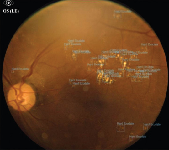
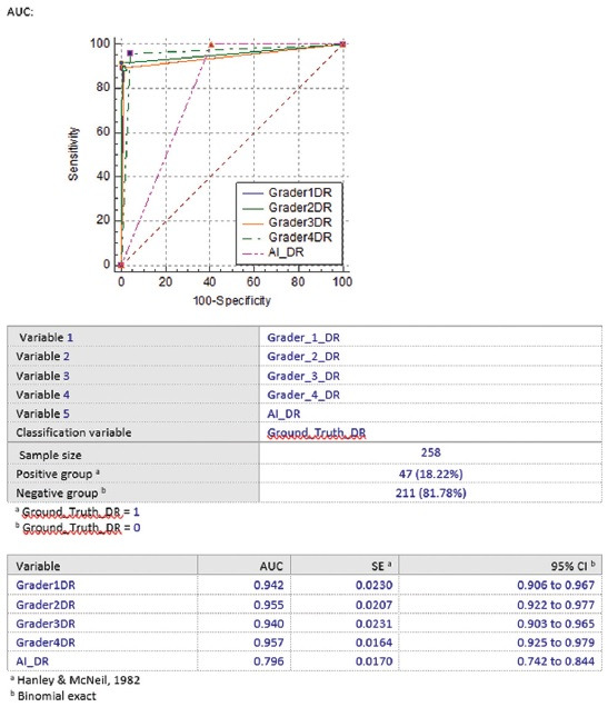
Methods: This cross-sectional study was carried out among 138 patients who underwent routine ophthalmic examination, irrespective of their diabetic status. The participants were subjected to a single field color fundus photography using nonmydriatic fundus camera. The images acquired were processed by AI algorithm for image quality, presence and refer ability of DR. The results were graded by four ophthalmologists. Interobserver variability between the four observers was also calculated.
Results: Of the 138 patients, 26 patients (18.84%) had some stage of DR, represented by 47 images (17.03%) positive for signs of DR. All 26 patients were immoderate or severe stage. About 6.5% of the images were considered as not gradable due to poor optical quality. The average agreement between pairs of the four graders was 95.16% for referable DR (RDR). The AI showed 100% sensitivity in detecting DR while the specificity for RDR was 91.47%.
Conclusion: AI has shown excellent sensitivity and specificity in RDR detection, at par with the performance of individual ophthalmologists and is an invaluable tool for DR screening.
期刊介绍:
The Middle East African Journal of Ophthalmology (MEAJO), published four times per year in print and online, is an official journal of the Middle East African Council of Ophthalmology (MEACO). It is an international, peer-reviewed journal whose mission includes publication of original research of interest to ophthalmologists in the Middle East and Africa, and to provide readers with high quality educational review articles from world-renown experts. MEAJO, previously known as Middle East Journal of Ophthalmology (MEJO) was founded by Dr Akef El Maghraby in 1993.

 求助内容:
求助内容: 应助结果提醒方式:


