Matti Sievert, Konstantinos Mantsopoulos, Sarina K Mueller, Markus Eckstein, Robin Rupp, Marc Aubreville, Florian Stelzle, Nicolai Oetter, Andreas Maier, Heinrich Iro, Miguel Goncalves
{"title":"共聚焦激光内镜的系统解释:喉和咽共聚焦成像评分。","authors":"Matti Sievert, Konstantinos Mantsopoulos, Sarina K Mueller, Markus Eckstein, Robin Rupp, Marc Aubreville, Florian Stelzle, Nicolai Oetter, Andreas Maier, Heinrich Iro, Miguel Goncalves","doi":"10.14639/0392-100X-N1643","DOIUrl":null,"url":null,"abstract":"<p><strong>Objective: </strong>Development and validation of a confocal laser endomicroscopy (CLE) classification score for the larynx and pharynx.</p><p><strong>Methods: </strong>Thirteen patients (154 video sequences, 9240 images) with laryngeal or pharyngeal SCC were included in this prospective study between October 2020 and February 2021. Each CLE sequence was correlated with the gold standard of histopathological examination. Based on a dataset of 94 video sequences (5640 images), a scoring system was developed. In the remaining 60 sequences (3600 images), the score was validated by four CLE experts and four head and neck surgeons who were not familiar with CLE.</p><p><strong>Results: </strong>Tissue homogeneity, cell size, borders and clusters, capillary loops and the nucleus/cytoplasm ratio were defined as the scoring criteria. Using this score, the CLE experts obtained an accuracy, sensitivity, and specificity of 90.8%, 95.1%, and 86.4%, respectively, and the CLE non-experts of 86.2%, 86.4%, and 86.1%. Interobserver agreement Fleiss' kappa was 0.8 and 0.6, respectively.</p><p><strong>Conclusions: </strong>CLE can be reliably evaluated based on defined and reproducible imaging features, which demonstrate a high diagnostic value. CLE can be easily integrated into the intraoperative setting and generate real-time, in-vivo microscopic images to demarcate malignant changes.</p>","PeriodicalId":520544,"journal":{"name":"Acta otorhinolaryngologica Italica : organo ufficiale della Societa italiana di otorinolaringologia e chirurgia cervico-facciale","volume":" ","pages":"26-33"},"PeriodicalIF":0.0000,"publicationDate":"2022-02-01","publicationTypes":"Journal Article","fieldsOfStudy":null,"isOpenAccess":false,"openAccessPdf":"https://ftp.ncbi.nlm.nih.gov/pub/pmc/oa_pdf/eb/6d/aoi-2022-01-26.PMC9058938.pdf","citationCount":"6","resultStr":"{\"title\":\"Systematic interpretation of confocal laser endomicroscopy: larynx and pharynx confocal imaging score.\",\"authors\":\"Matti Sievert, Konstantinos Mantsopoulos, Sarina K Mueller, Markus Eckstein, Robin Rupp, Marc Aubreville, Florian Stelzle, Nicolai Oetter, Andreas Maier, Heinrich Iro, Miguel Goncalves\",\"doi\":\"10.14639/0392-100X-N1643\",\"DOIUrl\":null,\"url\":null,\"abstract\":\"<p><strong>Objective: </strong>Development and validation of a confocal laser endomicroscopy (CLE) classification score for the larynx and pharynx.</p><p><strong>Methods: </strong>Thirteen patients (154 video sequences, 9240 images) with laryngeal or pharyngeal SCC were included in this prospective study between October 2020 and February 2021. Each CLE sequence was correlated with the gold standard of histopathological examination. Based on a dataset of 94 video sequences (5640 images), a scoring system was developed. In the remaining 60 sequences (3600 images), the score was validated by four CLE experts and four head and neck surgeons who were not familiar with CLE.</p><p><strong>Results: </strong>Tissue homogeneity, cell size, borders and clusters, capillary loops and the nucleus/cytoplasm ratio were defined as the scoring criteria. Using this score, the CLE experts obtained an accuracy, sensitivity, and specificity of 90.8%, 95.1%, and 86.4%, respectively, and the CLE non-experts of 86.2%, 86.4%, and 86.1%. Interobserver agreement Fleiss' kappa was 0.8 and 0.6, respectively.</p><p><strong>Conclusions: </strong>CLE can be reliably evaluated based on defined and reproducible imaging features, which demonstrate a high diagnostic value. CLE can be easily integrated into the intraoperative setting and generate real-time, in-vivo microscopic images to demarcate malignant changes.</p>\",\"PeriodicalId\":520544,\"journal\":{\"name\":\"Acta otorhinolaryngologica Italica : organo ufficiale della Societa italiana di otorinolaringologia e chirurgia cervico-facciale\",\"volume\":\" \",\"pages\":\"26-33\"},\"PeriodicalIF\":0.0000,\"publicationDate\":\"2022-02-01\",\"publicationTypes\":\"Journal Article\",\"fieldsOfStudy\":null,\"isOpenAccess\":false,\"openAccessPdf\":\"https://ftp.ncbi.nlm.nih.gov/pub/pmc/oa_pdf/eb/6d/aoi-2022-01-26.PMC9058938.pdf\",\"citationCount\":\"6\",\"resultStr\":null,\"platform\":\"Semanticscholar\",\"paperid\":null,\"PeriodicalName\":\"Acta otorhinolaryngologica Italica : organo ufficiale della Societa italiana di otorinolaringologia e chirurgia cervico-facciale\",\"FirstCategoryId\":\"3\",\"ListUrlMain\":\"https://doi.org/10.14639/0392-100X-N1643\",\"RegionNum\":0,\"RegionCategory\":null,\"ArticlePicture\":[],\"TitleCN\":null,\"AbstractTextCN\":null,\"PMCID\":null,\"EPubDate\":\"2022/2/7 0:00:00\",\"PubModel\":\"Epub\",\"JCR\":\"\",\"JCRName\":\"\",\"Score\":null,\"Total\":0}","platform":"Semanticscholar","paperid":null,"PeriodicalName":"Acta otorhinolaryngologica Italica : organo ufficiale della Societa italiana di otorinolaringologia e chirurgia cervico-facciale","FirstCategoryId":"3","ListUrlMain":"https://doi.org/10.14639/0392-100X-N1643","RegionNum":0,"RegionCategory":null,"ArticlePicture":[],"TitleCN":null,"AbstractTextCN":null,"PMCID":null,"EPubDate":"2022/2/7 0:00:00","PubModel":"Epub","JCR":"","JCRName":"","Score":null,"Total":0}
Systematic interpretation of confocal laser endomicroscopy: larynx and pharynx confocal imaging score.
Objective: Development and validation of a confocal laser endomicroscopy (CLE) classification score for the larynx and pharynx.
Methods: Thirteen patients (154 video sequences, 9240 images) with laryngeal or pharyngeal SCC were included in this prospective study between October 2020 and February 2021. Each CLE sequence was correlated with the gold standard of histopathological examination. Based on a dataset of 94 video sequences (5640 images), a scoring system was developed. In the remaining 60 sequences (3600 images), the score was validated by four CLE experts and four head and neck surgeons who were not familiar with CLE.
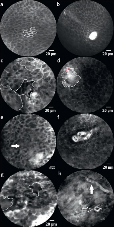
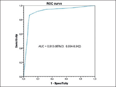
Results: Tissue homogeneity, cell size, borders and clusters, capillary loops and the nucleus/cytoplasm ratio were defined as the scoring criteria. Using this score, the CLE experts obtained an accuracy, sensitivity, and specificity of 90.8%, 95.1%, and 86.4%, respectively, and the CLE non-experts of 86.2%, 86.4%, and 86.1%. Interobserver agreement Fleiss' kappa was 0.8 and 0.6, respectively.
Conclusions: CLE can be reliably evaluated based on defined and reproducible imaging features, which demonstrate a high diagnostic value. CLE can be easily integrated into the intraoperative setting and generate real-time, in-vivo microscopic images to demarcate malignant changes.

 求助内容:
求助内容: 应助结果提醒方式:


