Juan D Arias, Francisco J Arango, Maria Margarita Parra, Ronald M Sánchez-Ávila, Gustavo A Parra-Serrano, Andrea T Hoyos, Silvia J Granados, Eduardo J Viteri, Ivetteh Gaibor-Santos, Yanny Perez
{"title":"光学相干断层扫描血管造影评价糖尿病前期患者早期微血管变化。","authors":"Juan D Arias, Francisco J Arango, Maria Margarita Parra, Ronald M Sánchez-Ávila, Gustavo A Parra-Serrano, Andrea T Hoyos, Silvia J Granados, Eduardo J Viteri, Ivetteh Gaibor-Santos, Yanny Perez","doi":"10.1177/25158414211047020","DOIUrl":null,"url":null,"abstract":"<p><strong>Background: </strong>Timely detection of early microvascular changes in patients with prediabetes could help reduce the likelihood of progression of diabetes-related retinal complications.</p><p><strong>Aim: </strong>To determine early microvascular changes in patients with prediabetes using optical coherence tomography angiography (OCT-A).</p><p><strong>Methods: </strong>In this single-center retrospective case-control study, macular OCT-A images of superficial capillary plexus (SCP) and deep capillary plexus (DCP) were analyzed in non-diabetic controls, and prediabetic and diabetic subjects. A quantitative analysis was performed using ImageJ software of the foveal avascular zone (FAZ) area, acircularity index (AI), perfusion density (PD), and vascular length density (VLD).</p><p><strong>Results: </strong>A total of 94 eyes of 53 patients were included in this study. The global mean age was 57.7 years, 39.6% men and 60.4% women. In SCP, the mean PD was 0.283 ± 0.15, 0.186 ± 0.720, and 0.186 ± 0.07 in non-diabetic controls, and prediabetic and diabetic groups, respectively. The mean VLD was 8.728 ± 3.425 in non-diabetic controls, 6.147 ± 1.399 in prediabetic group, and 6.292 ± 1.997 in patients with diabetes. The comparison of prediabetic patients and controls shows statistical differences between PD and VLD in both plexus SCP (<i>p</i> = 0.002 and <i>p</i> = 0.001, respectively) and DCP (<i>p</i> = 0.005 and <i>p</i> = 0.002, respectively). The mean area of FAZ in patients with diabetes and normal individuals was 0.281 and 0.196 mm<sup>2</sup>, respectively (<i>p</i> < 0.001). AI was higher in the control group (0.87 ± 0.14) and prediabetic group (0.80 ± 0.17) compared to diabetic patients (0.64 ± 0.19). There were no differences in FAZ area and AI between prediabetic and non-diabetic controls.</p><p><strong>Conclusion: </strong>PD and VLD demonstrated to be early microvascular changes in prediabetic patients evaluated by OCT-A. No alterations of FAZ were evidenced in this group.</p>","PeriodicalId":23054,"journal":{"name":"Therapeutic Advances in Ophthalmology","volume":"13 ","pages":"25158414211047020"},"PeriodicalIF":2.3000,"publicationDate":"2021-10-21","publicationTypes":"Journal Article","fieldsOfStudy":null,"isOpenAccess":false,"openAccessPdf":"https://ftp.ncbi.nlm.nih.gov/pub/pmc/oa_pdf/b3/e9/10.1177_25158414211047020.PMC8543708.pdf","citationCount":"4","resultStr":"{\"title\":\"Early microvascular changes in patients with prediabetes evaluated by optical coherence tomography angiography.\",\"authors\":\"Juan D Arias, Francisco J Arango, Maria Margarita Parra, Ronald M Sánchez-Ávila, Gustavo A Parra-Serrano, Andrea T Hoyos, Silvia J Granados, Eduardo J Viteri, Ivetteh Gaibor-Santos, Yanny Perez\",\"doi\":\"10.1177/25158414211047020\",\"DOIUrl\":null,\"url\":null,\"abstract\":\"<p><strong>Background: </strong>Timely detection of early microvascular changes in patients with prediabetes could help reduce the likelihood of progression of diabetes-related retinal complications.</p><p><strong>Aim: </strong>To determine early microvascular changes in patients with prediabetes using optical coherence tomography angiography (OCT-A).</p><p><strong>Methods: </strong>In this single-center retrospective case-control study, macular OCT-A images of superficial capillary plexus (SCP) and deep capillary plexus (DCP) were analyzed in non-diabetic controls, and prediabetic and diabetic subjects. A quantitative analysis was performed using ImageJ software of the foveal avascular zone (FAZ) area, acircularity index (AI), perfusion density (PD), and vascular length density (VLD).</p><p><strong>Results: </strong>A total of 94 eyes of 53 patients were included in this study. The global mean age was 57.7 years, 39.6% men and 60.4% women. In SCP, the mean PD was 0.283 ± 0.15, 0.186 ± 0.720, and 0.186 ± 0.07 in non-diabetic controls, and prediabetic and diabetic groups, respectively. The mean VLD was 8.728 ± 3.425 in non-diabetic controls, 6.147 ± 1.399 in prediabetic group, and 6.292 ± 1.997 in patients with diabetes. The comparison of prediabetic patients and controls shows statistical differences between PD and VLD in both plexus SCP (<i>p</i> = 0.002 and <i>p</i> = 0.001, respectively) and DCP (<i>p</i> = 0.005 and <i>p</i> = 0.002, respectively). The mean area of FAZ in patients with diabetes and normal individuals was 0.281 and 0.196 mm<sup>2</sup>, respectively (<i>p</i> < 0.001). AI was higher in the control group (0.87 ± 0.14) and prediabetic group (0.80 ± 0.17) compared to diabetic patients (0.64 ± 0.19). There were no differences in FAZ area and AI between prediabetic and non-diabetic controls.</p><p><strong>Conclusion: </strong>PD and VLD demonstrated to be early microvascular changes in prediabetic patients evaluated by OCT-A. No alterations of FAZ were evidenced in this group.</p>\",\"PeriodicalId\":23054,\"journal\":{\"name\":\"Therapeutic Advances in Ophthalmology\",\"volume\":\"13 \",\"pages\":\"25158414211047020\"},\"PeriodicalIF\":2.3000,\"publicationDate\":\"2021-10-21\",\"publicationTypes\":\"Journal Article\",\"fieldsOfStudy\":null,\"isOpenAccess\":false,\"openAccessPdf\":\"https://ftp.ncbi.nlm.nih.gov/pub/pmc/oa_pdf/b3/e9/10.1177_25158414211047020.PMC8543708.pdf\",\"citationCount\":\"4\",\"resultStr\":null,\"platform\":\"Semanticscholar\",\"paperid\":null,\"PeriodicalName\":\"Therapeutic Advances in Ophthalmology\",\"FirstCategoryId\":\"1085\",\"ListUrlMain\":\"https://doi.org/10.1177/25158414211047020\",\"RegionNum\":0,\"RegionCategory\":null,\"ArticlePicture\":[],\"TitleCN\":null,\"AbstractTextCN\":null,\"PMCID\":null,\"EPubDate\":\"2021/1/1 0:00:00\",\"PubModel\":\"eCollection\",\"JCR\":\"Q2\",\"JCRName\":\"OPHTHALMOLOGY\",\"Score\":null,\"Total\":0}","platform":"Semanticscholar","paperid":null,"PeriodicalName":"Therapeutic Advances in Ophthalmology","FirstCategoryId":"1085","ListUrlMain":"https://doi.org/10.1177/25158414211047020","RegionNum":0,"RegionCategory":null,"ArticlePicture":[],"TitleCN":null,"AbstractTextCN":null,"PMCID":null,"EPubDate":"2021/1/1 0:00:00","PubModel":"eCollection","JCR":"Q2","JCRName":"OPHTHALMOLOGY","Score":null,"Total":0}
Early microvascular changes in patients with prediabetes evaluated by optical coherence tomography angiography.
Background: Timely detection of early microvascular changes in patients with prediabetes could help reduce the likelihood of progression of diabetes-related retinal complications.
Aim: To determine early microvascular changes in patients with prediabetes using optical coherence tomography angiography (OCT-A).
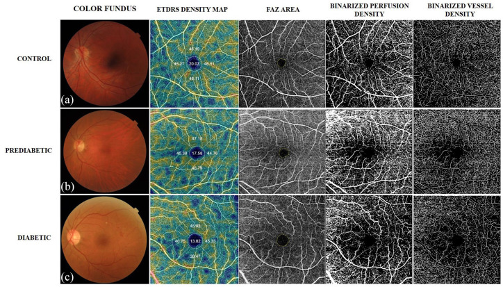
Methods: In this single-center retrospective case-control study, macular OCT-A images of superficial capillary plexus (SCP) and deep capillary plexus (DCP) were analyzed in non-diabetic controls, and prediabetic and diabetic subjects. A quantitative analysis was performed using ImageJ software of the foveal avascular zone (FAZ) area, acircularity index (AI), perfusion density (PD), and vascular length density (VLD).
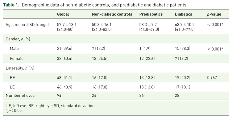
Results: A total of 94 eyes of 53 patients were included in this study. The global mean age was 57.7 years, 39.6% men and 60.4% women. In SCP, the mean PD was 0.283 ± 0.15, 0.186 ± 0.720, and 0.186 ± 0.07 in non-diabetic controls, and prediabetic and diabetic groups, respectively. The mean VLD was 8.728 ± 3.425 in non-diabetic controls, 6.147 ± 1.399 in prediabetic group, and 6.292 ± 1.997 in patients with diabetes. The comparison of prediabetic patients and controls shows statistical differences between PD and VLD in both plexus SCP (p = 0.002 and p = 0.001, respectively) and DCP (p = 0.005 and p = 0.002, respectively). The mean area of FAZ in patients with diabetes and normal individuals was 0.281 and 0.196 mm2, respectively (p < 0.001). AI was higher in the control group (0.87 ± 0.14) and prediabetic group (0.80 ± 0.17) compared to diabetic patients (0.64 ± 0.19). There were no differences in FAZ area and AI between prediabetic and non-diabetic controls.
Conclusion: PD and VLD demonstrated to be early microvascular changes in prediabetic patients evaluated by OCT-A. No alterations of FAZ were evidenced in this group.


 求助内容:
求助内容: 应助结果提醒方式:


