Fabienne Paschereit, Kim Hannah Schindelmann, Michael Hummel, Joanna Schneider, Gisela Stoltenburg-Didinger, Angela M Kaindl
{"title":"脊柱裂的脑异常:一项神经病理学研究。","authors":"Fabienne Paschereit, Kim Hannah Schindelmann, Michael Hummel, Joanna Schneider, Gisela Stoltenburg-Didinger, Angela M Kaindl","doi":"10.1177/10935266211040500","DOIUrl":null,"url":null,"abstract":"<p><strong>Introduction: </strong>Spina bifida (SB) is the most common neural tube defect in humans. Here, we analyzed systematically the neuropathological findings of the brain in SB cases.</p><p><strong>Methods: </strong>79 cases with SB aperta (SBA) and 6 cases with SB occulta (SBO) autopsied at the Charité Neuropathology from 1974 to 2000 were re-evaluated retrospectively. For this, case files and spinal cord as well as brain sections were studied.</p><p><strong>Results: </strong>While no brain malformations were detected in SBO cases, 95% of SBA cases had brain malformations. Main brain anomalies identified were hydrocephalus (71%), Chiari II malformation (36%), heterotopia (34%), other cerebellar anomalies (36%), gyrification defects (33%), and ependymal denudation (29%). Hydrocephalus was observed as early as gestational week 17 and was highly associated to Chiari II and ependymal denudation. In 55% SBA was accompanied by further anomalies not primarily affecting the CNS.</p><p><strong>Conclusion: </strong>We confirm using neuropathologic methods brain malformations in most SBA but none in SBO cases. In addition to our previous radiologic study, we now demonstrate the high prevalence of cerebellar malformations and cerebral heterotopias in SBA. The early detection of hydrocephalus and Chiari II malformation in fetuses raises the question whether these arise parallel rather than in strict temporal sequence.</p>","PeriodicalId":520743,"journal":{"name":"Pediatric and developmental pathology : the official journal of the Society for Pediatric Pathology and the Paediatric Pathology Society","volume":" ","pages":"107-123"},"PeriodicalIF":1.3000,"publicationDate":"2022-03-01","publicationTypes":"Journal Article","fieldsOfStudy":null,"isOpenAccess":false,"openAccessPdf":"https://ftp.ncbi.nlm.nih.gov/pub/pmc/oa_pdf/d4/7e/10.1177_10935266211040500.PMC9109215.pdf","citationCount":"0","resultStr":"{\"title\":\"Cerebral Abnormalities in Spina Bifida: A Neuropathological Study.\",\"authors\":\"Fabienne Paschereit, Kim Hannah Schindelmann, Michael Hummel, Joanna Schneider, Gisela Stoltenburg-Didinger, Angela M Kaindl\",\"doi\":\"10.1177/10935266211040500\",\"DOIUrl\":null,\"url\":null,\"abstract\":\"<p><strong>Introduction: </strong>Spina bifida (SB) is the most common neural tube defect in humans. Here, we analyzed systematically the neuropathological findings of the brain in SB cases.</p><p><strong>Methods: </strong>79 cases with SB aperta (SBA) and 6 cases with SB occulta (SBO) autopsied at the Charité Neuropathology from 1974 to 2000 were re-evaluated retrospectively. For this, case files and spinal cord as well as brain sections were studied.</p><p><strong>Results: </strong>While no brain malformations were detected in SBO cases, 95% of SBA cases had brain malformations. Main brain anomalies identified were hydrocephalus (71%), Chiari II malformation (36%), heterotopia (34%), other cerebellar anomalies (36%), gyrification defects (33%), and ependymal denudation (29%). Hydrocephalus was observed as early as gestational week 17 and was highly associated to Chiari II and ependymal denudation. In 55% SBA was accompanied by further anomalies not primarily affecting the CNS.</p><p><strong>Conclusion: </strong>We confirm using neuropathologic methods brain malformations in most SBA but none in SBO cases. In addition to our previous radiologic study, we now demonstrate the high prevalence of cerebellar malformations and cerebral heterotopias in SBA. The early detection of hydrocephalus and Chiari II malformation in fetuses raises the question whether these arise parallel rather than in strict temporal sequence.</p>\",\"PeriodicalId\":520743,\"journal\":{\"name\":\"Pediatric and developmental pathology : the official journal of the Society for Pediatric Pathology and the Paediatric Pathology Society\",\"volume\":\" \",\"pages\":\"107-123\"},\"PeriodicalIF\":1.3000,\"publicationDate\":\"2022-03-01\",\"publicationTypes\":\"Journal Article\",\"fieldsOfStudy\":null,\"isOpenAccess\":false,\"openAccessPdf\":\"https://ftp.ncbi.nlm.nih.gov/pub/pmc/oa_pdf/d4/7e/10.1177_10935266211040500.PMC9109215.pdf\",\"citationCount\":\"0\",\"resultStr\":null,\"platform\":\"Semanticscholar\",\"paperid\":null,\"PeriodicalName\":\"Pediatric and developmental pathology : the official journal of the Society for Pediatric Pathology and the Paediatric Pathology Society\",\"FirstCategoryId\":\"3\",\"ListUrlMain\":\"https://doi.org/10.1177/10935266211040500\",\"RegionNum\":0,\"RegionCategory\":null,\"ArticlePicture\":[],\"TitleCN\":null,\"AbstractTextCN\":null,\"PMCID\":null,\"EPubDate\":\"2021/10/6 0:00:00\",\"PubModel\":\"Epub\",\"JCR\":\"\",\"JCRName\":\"\",\"Score\":null,\"Total\":0}","platform":"Semanticscholar","paperid":null,"PeriodicalName":"Pediatric and developmental pathology : the official journal of the Society for Pediatric Pathology and the Paediatric Pathology Society","FirstCategoryId":"3","ListUrlMain":"https://doi.org/10.1177/10935266211040500","RegionNum":0,"RegionCategory":null,"ArticlePicture":[],"TitleCN":null,"AbstractTextCN":null,"PMCID":null,"EPubDate":"2021/10/6 0:00:00","PubModel":"Epub","JCR":"","JCRName":"","Score":null,"Total":0}
Cerebral Abnormalities in Spina Bifida: A Neuropathological Study.
Introduction: Spina bifida (SB) is the most common neural tube defect in humans. Here, we analyzed systematically the neuropathological findings of the brain in SB cases.
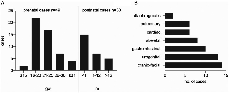
Methods: 79 cases with SB aperta (SBA) and 6 cases with SB occulta (SBO) autopsied at the Charité Neuropathology from 1974 to 2000 were re-evaluated retrospectively. For this, case files and spinal cord as well as brain sections were studied.
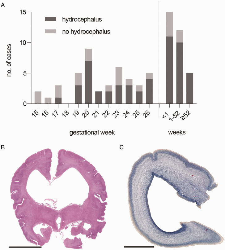
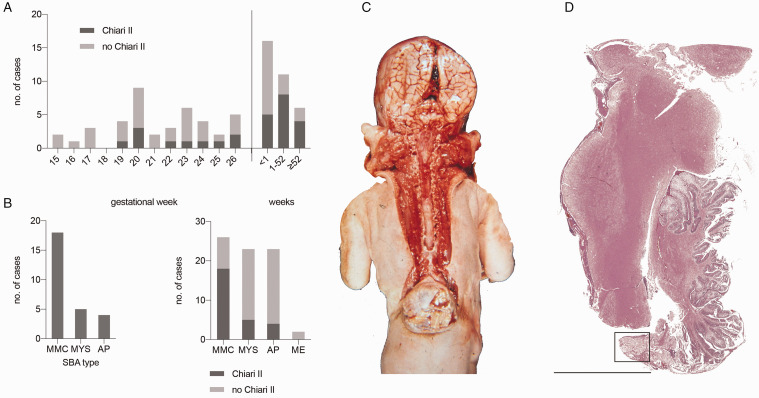
Results: While no brain malformations were detected in SBO cases, 95% of SBA cases had brain malformations. Main brain anomalies identified were hydrocephalus (71%), Chiari II malformation (36%), heterotopia (34%), other cerebellar anomalies (36%), gyrification defects (33%), and ependymal denudation (29%). Hydrocephalus was observed as early as gestational week 17 and was highly associated to Chiari II and ependymal denudation. In 55% SBA was accompanied by further anomalies not primarily affecting the CNS.
Conclusion: We confirm using neuropathologic methods brain malformations in most SBA but none in SBO cases. In addition to our previous radiologic study, we now demonstrate the high prevalence of cerebellar malformations and cerebral heterotopias in SBA. The early detection of hydrocephalus and Chiari II malformation in fetuses raises the question whether these arise parallel rather than in strict temporal sequence.

 求助内容:
求助内容: 应助结果提醒方式:


