Waldo Sepulveda, Francisco Sepulveda, Gloria Gonzalez, Claudio Arce, Elisa Alcalde
{"title":"先天性肝囊肿:产前和产后影像学表现。","authors":"Waldo Sepulveda, Francisco Sepulveda, Gloria Gonzalez, Claudio Arce, Elisa Alcalde","doi":"10.1177/1742271X20970601","DOIUrl":null,"url":null,"abstract":"<p><strong>Introduction: </strong>Congenital hepatic cyst is a rare hepatobiliary malformation that can present as an asymptomatic, unilocular, upper abdominal cystic mass in the fetus.</p><p><strong>Cases: </strong>We report two cases of congenital hepatic cyst in which the diagnosis was made by prenatal ultrasound at 25 and 33 weeks' gestation. The diagnosis was confirmed postnatally by abdominal ultrasound and radiologic imaging studies. Although the infants remained asymptomatic, laparoscopic excision was performed due to the increasing size of the cyst in both cases. Pathological examination of the resected specimens confirmed a simple cyst in one case and an epidermoid cyst in the other.</p><p><strong>Conclusions: </strong>Our cases and those described in the literature demonstrate the usefulness of incidental prenatal detection of congenital hepatic cyst, especially during late pregnancy. Such a diagnosis can allow for proper perinatal surveillance, selection of the route of delivery, and timely postnatal surgical intervention if required.</p>","PeriodicalId":23440,"journal":{"name":"Ultrasound","volume":"29 3","pages":"193-198"},"PeriodicalIF":0.8000,"publicationDate":"2021-08-01","publicationTypes":"Journal Article","fieldsOfStudy":null,"isOpenAccess":false,"openAccessPdf":"https://sci-hub-pdf.com/10.1177/1742271X20970601","citationCount":"4","resultStr":"{\"title\":\"Congenital hepatic cyst: Prenatal and postnatal imaging findings.\",\"authors\":\"Waldo Sepulveda, Francisco Sepulveda, Gloria Gonzalez, Claudio Arce, Elisa Alcalde\",\"doi\":\"10.1177/1742271X20970601\",\"DOIUrl\":null,\"url\":null,\"abstract\":\"<p><strong>Introduction: </strong>Congenital hepatic cyst is a rare hepatobiliary malformation that can present as an asymptomatic, unilocular, upper abdominal cystic mass in the fetus.</p><p><strong>Cases: </strong>We report two cases of congenital hepatic cyst in which the diagnosis was made by prenatal ultrasound at 25 and 33 weeks' gestation. The diagnosis was confirmed postnatally by abdominal ultrasound and radiologic imaging studies. Although the infants remained asymptomatic, laparoscopic excision was performed due to the increasing size of the cyst in both cases. Pathological examination of the resected specimens confirmed a simple cyst in one case and an epidermoid cyst in the other.</p><p><strong>Conclusions: </strong>Our cases and those described in the literature demonstrate the usefulness of incidental prenatal detection of congenital hepatic cyst, especially during late pregnancy. Such a diagnosis can allow for proper perinatal surveillance, selection of the route of delivery, and timely postnatal surgical intervention if required.</p>\",\"PeriodicalId\":23440,\"journal\":{\"name\":\"Ultrasound\",\"volume\":\"29 3\",\"pages\":\"193-198\"},\"PeriodicalIF\":0.8000,\"publicationDate\":\"2021-08-01\",\"publicationTypes\":\"Journal Article\",\"fieldsOfStudy\":null,\"isOpenAccess\":false,\"openAccessPdf\":\"https://sci-hub-pdf.com/10.1177/1742271X20970601\",\"citationCount\":\"4\",\"resultStr\":null,\"platform\":\"Semanticscholar\",\"paperid\":null,\"PeriodicalName\":\"Ultrasound\",\"FirstCategoryId\":\"1085\",\"ListUrlMain\":\"https://doi.org/10.1177/1742271X20970601\",\"RegionNum\":0,\"RegionCategory\":null,\"ArticlePicture\":[],\"TitleCN\":null,\"AbstractTextCN\":null,\"PMCID\":null,\"EPubDate\":\"2020/11/13 0:00:00\",\"PubModel\":\"Epub\",\"JCR\":\"Q4\",\"JCRName\":\"RADIOLOGY, NUCLEAR MEDICINE & MEDICAL IMAGING\",\"Score\":null,\"Total\":0}","platform":"Semanticscholar","paperid":null,"PeriodicalName":"Ultrasound","FirstCategoryId":"1085","ListUrlMain":"https://doi.org/10.1177/1742271X20970601","RegionNum":0,"RegionCategory":null,"ArticlePicture":[],"TitleCN":null,"AbstractTextCN":null,"PMCID":null,"EPubDate":"2020/11/13 0:00:00","PubModel":"Epub","JCR":"Q4","JCRName":"RADIOLOGY, NUCLEAR MEDICINE & MEDICAL IMAGING","Score":null,"Total":0}
Congenital hepatic cyst: Prenatal and postnatal imaging findings.
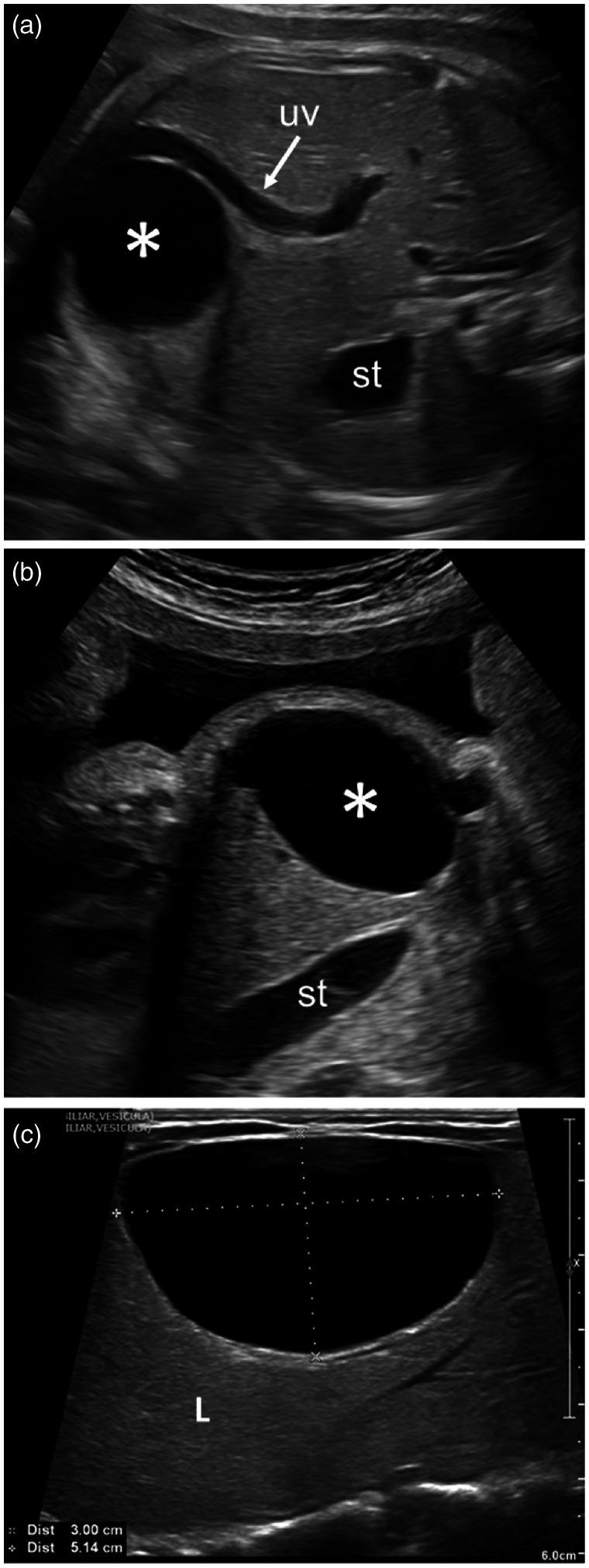
Introduction: Congenital hepatic cyst is a rare hepatobiliary malformation that can present as an asymptomatic, unilocular, upper abdominal cystic mass in the fetus.
Cases: We report two cases of congenital hepatic cyst in which the diagnosis was made by prenatal ultrasound at 25 and 33 weeks' gestation. The diagnosis was confirmed postnatally by abdominal ultrasound and radiologic imaging studies. Although the infants remained asymptomatic, laparoscopic excision was performed due to the increasing size of the cyst in both cases. Pathological examination of the resected specimens confirmed a simple cyst in one case and an epidermoid cyst in the other.
Conclusions: Our cases and those described in the literature demonstrate the usefulness of incidental prenatal detection of congenital hepatic cyst, especially during late pregnancy. Such a diagnosis can allow for proper perinatal surveillance, selection of the route of delivery, and timely postnatal surgical intervention if required.
UltrasoundRADIOLOGY, NUCLEAR MEDICINE & MEDICAL IMAGING-
CiteScore
1.70
自引率
0.00%
发文量
55
期刊介绍:
Ultrasound is the official journal of the British Medical Ultrasound Society (BMUS), a multidisciplinary, charitable society comprising radiologists, obstetricians, sonographers, physicists and veterinarians amongst others.

 求助内容:
求助内容: 应助结果提醒方式:


