A A Korytkin, N Yu Orlinskaya, Ya S Novikova, S A Gerasimov, D V Davydenko, K V Kulakova, S I Tverdokhlebov, E N Bolbasov
{"title":"不同孔隙度磷酸钙包覆与非包覆钛种植体的生物相容性与骨整合。","authors":"A A Korytkin, N Yu Orlinskaya, Ya S Novikova, S A Gerasimov, D V Davydenko, K V Kulakova, S I Tverdokhlebov, E N Bolbasov","doi":"10.17691/stm2021.13.2.06","DOIUrl":null,"url":null,"abstract":"<p><p><b>The aim of the investigation</b> was to study the influence of pore size and the presence of a biologically active calcium phosphate coating in porous 3D printed titanium implants on the process of integration with the bone tissue.</p><p><strong>Materials and methods: </strong>Samples of cylindrical implants with three different pore diameters (100, 200, and 400 μm) were fabricated from titanium powder on the Arcam 3D printer (Sweden) using electron beam melting technology. A calcium phosphate coating with a thickness of 20±4 μm was applied to some of the products by microarc oxidation. Cytotoxicity of the implants was determined <i>in vitro</i> on human dermal fibroblast cultures. The samples were implanted in the femoral bones of 36 rabbits <i>in vivo</i>. The animals were divided into 6 groups according to the bone implant samples. The prepared samples and peri-implant tissues were studied on days 90 and 180 after implantation using scanning electron microscopy and histological methods.</p><p><strong>Results: </strong>All samples under study were found to be non-toxic and well biocompatible with the bone tissue. There were revealed no differences between coated and non-coated implants of 100 and 200 μm pore diameters in terms of their histological structure, intensity of vascularization in the early stages, and bone formation in the later stages. Samples with pore diameters of 100 and 200 μm were easily removed from the bone tissue, the depth of bone growth into the pores of the implant was lower than in the samples with pore diameter of 400 μm (p<0.001). There were differences between coated and non-coated samples of 400 μm pore diameter, which was expressed in a more intensive osseointegration of samples with calcium phosphate coating (p<0.05).</p><p><strong>Conclusion: </strong>The optimal surface characteristics of the material for repairing bone defects are a pore diameter of 400 μm and the presence of a calcium phosphate coating.</p>","PeriodicalId":51886,"journal":{"name":"Sovremennye Tehnologii v Medicine","volume":"13 2","pages":"52-57"},"PeriodicalIF":0.9000,"publicationDate":"2021-01-01","publicationTypes":"Journal Article","fieldsOfStudy":null,"isOpenAccess":false,"openAccessPdf":"https://www.ncbi.nlm.nih.gov/pmc/articles/PMC8353716/pdf/","citationCount":"2","resultStr":"{\"title\":\"Biocompatibility and Osseointegration of Calcium Phosphate-Coated and Non-Coated Titanium Implants with Various Porosities.\",\"authors\":\"A A Korytkin, N Yu Orlinskaya, Ya S Novikova, S A Gerasimov, D V Davydenko, K V Kulakova, S I Tverdokhlebov, E N Bolbasov\",\"doi\":\"10.17691/stm2021.13.2.06\",\"DOIUrl\":null,\"url\":null,\"abstract\":\"<p><p><b>The aim of the investigation</b> was to study the influence of pore size and the presence of a biologically active calcium phosphate coating in porous 3D printed titanium implants on the process of integration with the bone tissue.</p><p><strong>Materials and methods: </strong>Samples of cylindrical implants with three different pore diameters (100, 200, and 400 μm) were fabricated from titanium powder on the Arcam 3D printer (Sweden) using electron beam melting technology. A calcium phosphate coating with a thickness of 20±4 μm was applied to some of the products by microarc oxidation. Cytotoxicity of the implants was determined <i>in vitro</i> on human dermal fibroblast cultures. The samples were implanted in the femoral bones of 36 rabbits <i>in vivo</i>. The animals were divided into 6 groups according to the bone implant samples. The prepared samples and peri-implant tissues were studied on days 90 and 180 after implantation using scanning electron microscopy and histological methods.</p><p><strong>Results: </strong>All samples under study were found to be non-toxic and well biocompatible with the bone tissue. There were revealed no differences between coated and non-coated implants of 100 and 200 μm pore diameters in terms of their histological structure, intensity of vascularization in the early stages, and bone formation in the later stages. Samples with pore diameters of 100 and 200 μm were easily removed from the bone tissue, the depth of bone growth into the pores of the implant was lower than in the samples with pore diameter of 400 μm (p<0.001). There were differences between coated and non-coated samples of 400 μm pore diameter, which was expressed in a more intensive osseointegration of samples with calcium phosphate coating (p<0.05).</p><p><strong>Conclusion: </strong>The optimal surface characteristics of the material for repairing bone defects are a pore diameter of 400 μm and the presence of a calcium phosphate coating.</p>\",\"PeriodicalId\":51886,\"journal\":{\"name\":\"Sovremennye Tehnologii v Medicine\",\"volume\":\"13 2\",\"pages\":\"52-57\"},\"PeriodicalIF\":0.9000,\"publicationDate\":\"2021-01-01\",\"publicationTypes\":\"Journal Article\",\"fieldsOfStudy\":null,\"isOpenAccess\":false,\"openAccessPdf\":\"https://www.ncbi.nlm.nih.gov/pmc/articles/PMC8353716/pdf/\",\"citationCount\":\"2\",\"resultStr\":null,\"platform\":\"Semanticscholar\",\"paperid\":null,\"PeriodicalName\":\"Sovremennye Tehnologii v Medicine\",\"FirstCategoryId\":\"1085\",\"ListUrlMain\":\"https://doi.org/10.17691/stm2021.13.2.06\",\"RegionNum\":0,\"RegionCategory\":null,\"ArticlePicture\":[],\"TitleCN\":null,\"AbstractTextCN\":null,\"PMCID\":null,\"EPubDate\":\"\",\"PubModel\":\"\",\"JCR\":\"Q4\",\"JCRName\":\"MEDICINE, RESEARCH & EXPERIMENTAL\",\"Score\":null,\"Total\":0}","platform":"Semanticscholar","paperid":null,"PeriodicalName":"Sovremennye Tehnologii v Medicine","FirstCategoryId":"1085","ListUrlMain":"https://doi.org/10.17691/stm2021.13.2.06","RegionNum":0,"RegionCategory":null,"ArticlePicture":[],"TitleCN":null,"AbstractTextCN":null,"PMCID":null,"EPubDate":"","PubModel":"","JCR":"Q4","JCRName":"MEDICINE, RESEARCH & EXPERIMENTAL","Score":null,"Total":0}
Biocompatibility and Osseointegration of Calcium Phosphate-Coated and Non-Coated Titanium Implants with Various Porosities.
The aim of the investigation was to study the influence of pore size and the presence of a biologically active calcium phosphate coating in porous 3D printed titanium implants on the process of integration with the bone tissue.
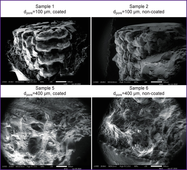
Materials and methods: Samples of cylindrical implants with three different pore diameters (100, 200, and 400 μm) were fabricated from titanium powder on the Arcam 3D printer (Sweden) using electron beam melting technology. A calcium phosphate coating with a thickness of 20±4 μm was applied to some of the products by microarc oxidation. Cytotoxicity of the implants was determined in vitro on human dermal fibroblast cultures. The samples were implanted in the femoral bones of 36 rabbits in vivo. The animals were divided into 6 groups according to the bone implant samples. The prepared samples and peri-implant tissues were studied on days 90 and 180 after implantation using scanning electron microscopy and histological methods.
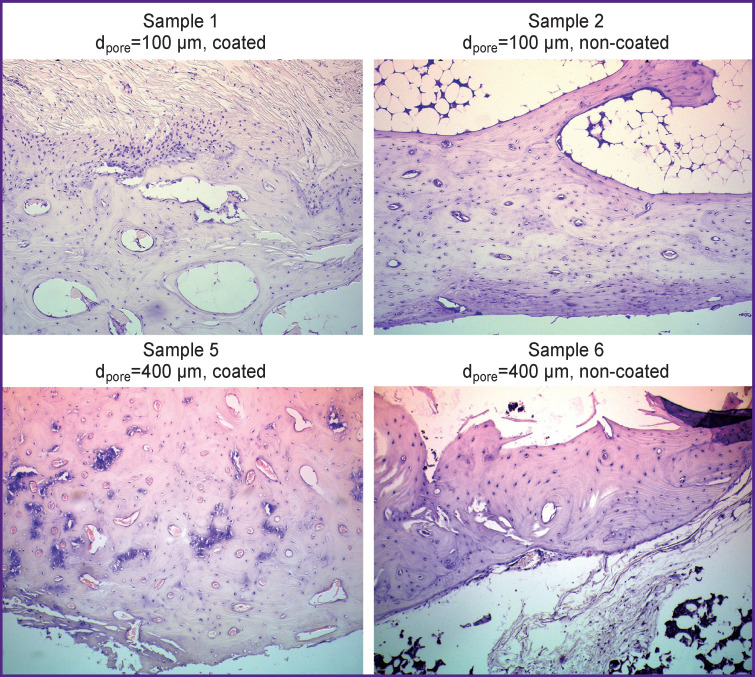
Results: All samples under study were found to be non-toxic and well biocompatible with the bone tissue. There were revealed no differences between coated and non-coated implants of 100 and 200 μm pore diameters in terms of their histological structure, intensity of vascularization in the early stages, and bone formation in the later stages. Samples with pore diameters of 100 and 200 μm were easily removed from the bone tissue, the depth of bone growth into the pores of the implant was lower than in the samples with pore diameter of 400 μm (p<0.001). There were differences between coated and non-coated samples of 400 μm pore diameter, which was expressed in a more intensive osseointegration of samples with calcium phosphate coating (p<0.05).
Conclusion: The optimal surface characteristics of the material for repairing bone defects are a pore diameter of 400 μm and the presence of a calcium phosphate coating.

 求助内容:
求助内容: 应助结果提醒方式:


