{"title":"兴奋性突触后钙瞬态在感觉-运动神经元突触允许在培养中对突触强度进行数天的定量检查。","authors":"Tyler W Dunn, Wayne S Sossin","doi":"10.1101/lm.052639.120","DOIUrl":null,"url":null,"abstract":"<p><p>A more thorough description of the changes in synaptic strength underlying synaptic plasticity may be achieved with quantal resolution measurements at individual synaptic sites. Here, we demonstrate that by using a membrane targeted genetic calcium sensor, we can measure quantal synaptic events at the individual synaptic sites of <i>Aplysia</i> sensory neuron to motor neuron synaptic connections. These results show that synaptic strength is not evenly distributed between all contacts in these cultures, but dominated by multiquantal sites of synaptic contact, likely clusters of individual synaptic sites. Surprisingly, most synaptic contacts were not found opposite presynaptic varicosities, but instead at areas of pre- and postsynaptic contact with no visible thickening of membranes. The release probability, quantal size, and quantal content can be measured over days at individual synaptic contacts using this technique. Homosynaptic depression was accompanied by a reduction in release site probability, with no evidence of individual synaptic site silencing over the course of depression. This technique shows promise in being able to address outstanding questions in this system, including determining the synaptic changes that maintain long-term alterations in synaptic strength that underlie memory.</p>","PeriodicalId":520703,"journal":{"name":"Learning & memory (Cold Spring Harbor, N.Y.)","volume":" ","pages":"277-290"},"PeriodicalIF":2.2000,"publicationDate":"2021-08-16","publicationTypes":"Journal Article","fieldsOfStudy":null,"isOpenAccess":false,"openAccessPdf":"https://ftp.ncbi.nlm.nih.gov/pub/pmc/oa_pdf/ef/5d/LM052639Dun.PMC8372562.pdf","citationCount":"2","resultStr":"{\"title\":\"Excitatory postsynaptic calcium transients at <i>Aplysia</i> sensory-motor neuron synapses allow for quantal examination of synaptic strength over multiple days in culture.\",\"authors\":\"Tyler W Dunn, Wayne S Sossin\",\"doi\":\"10.1101/lm.052639.120\",\"DOIUrl\":null,\"url\":null,\"abstract\":\"<p><p>A more thorough description of the changes in synaptic strength underlying synaptic plasticity may be achieved with quantal resolution measurements at individual synaptic sites. Here, we demonstrate that by using a membrane targeted genetic calcium sensor, we can measure quantal synaptic events at the individual synaptic sites of <i>Aplysia</i> sensory neuron to motor neuron synaptic connections. These results show that synaptic strength is not evenly distributed between all contacts in these cultures, but dominated by multiquantal sites of synaptic contact, likely clusters of individual synaptic sites. Surprisingly, most synaptic contacts were not found opposite presynaptic varicosities, but instead at areas of pre- and postsynaptic contact with no visible thickening of membranes. The release probability, quantal size, and quantal content can be measured over days at individual synaptic contacts using this technique. Homosynaptic depression was accompanied by a reduction in release site probability, with no evidence of individual synaptic site silencing over the course of depression. This technique shows promise in being able to address outstanding questions in this system, including determining the synaptic changes that maintain long-term alterations in synaptic strength that underlie memory.</p>\",\"PeriodicalId\":520703,\"journal\":{\"name\":\"Learning & memory (Cold Spring Harbor, N.Y.)\",\"volume\":\" \",\"pages\":\"277-290\"},\"PeriodicalIF\":2.2000,\"publicationDate\":\"2021-08-16\",\"publicationTypes\":\"Journal Article\",\"fieldsOfStudy\":null,\"isOpenAccess\":false,\"openAccessPdf\":\"https://ftp.ncbi.nlm.nih.gov/pub/pmc/oa_pdf/ef/5d/LM052639Dun.PMC8372562.pdf\",\"citationCount\":\"2\",\"resultStr\":null,\"platform\":\"Semanticscholar\",\"paperid\":null,\"PeriodicalName\":\"Learning & memory (Cold Spring Harbor, N.Y.)\",\"FirstCategoryId\":\"3\",\"ListUrlMain\":\"https://doi.org/10.1101/lm.052639.120\",\"RegionNum\":0,\"RegionCategory\":null,\"ArticlePicture\":[],\"TitleCN\":null,\"AbstractTextCN\":null,\"PMCID\":null,\"EPubDate\":\"2021/9/1 0:00:00\",\"PubModel\":\"Print\",\"JCR\":\"\",\"JCRName\":\"\",\"Score\":null,\"Total\":0}","platform":"Semanticscholar","paperid":null,"PeriodicalName":"Learning & memory (Cold Spring Harbor, N.Y.)","FirstCategoryId":"3","ListUrlMain":"https://doi.org/10.1101/lm.052639.120","RegionNum":0,"RegionCategory":null,"ArticlePicture":[],"TitleCN":null,"AbstractTextCN":null,"PMCID":null,"EPubDate":"2021/9/1 0:00:00","PubModel":"Print","JCR":"","JCRName":"","Score":null,"Total":0}
Excitatory postsynaptic calcium transients at Aplysia sensory-motor neuron synapses allow for quantal examination of synaptic strength over multiple days in culture.
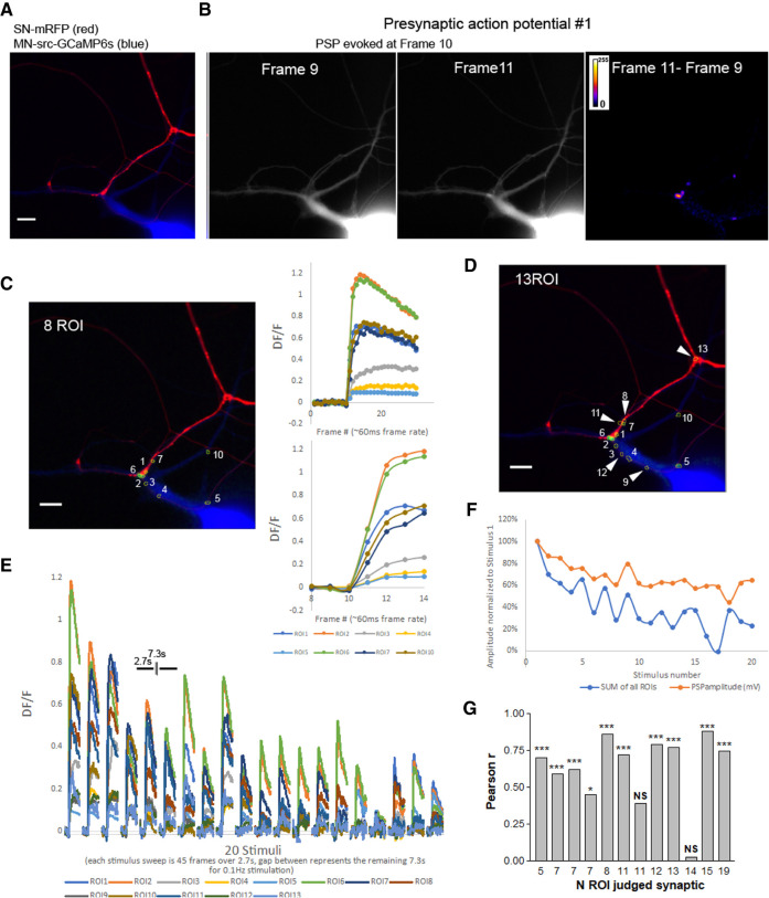
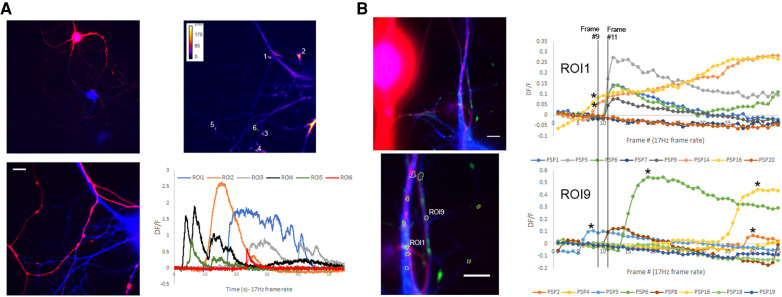
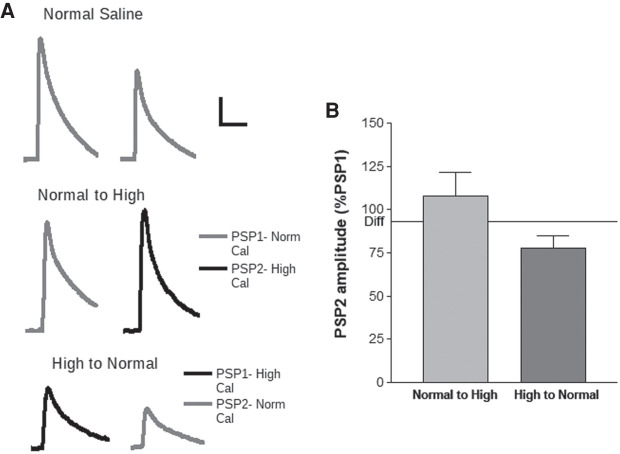
A more thorough description of the changes in synaptic strength underlying synaptic plasticity may be achieved with quantal resolution measurements at individual synaptic sites. Here, we demonstrate that by using a membrane targeted genetic calcium sensor, we can measure quantal synaptic events at the individual synaptic sites of Aplysia sensory neuron to motor neuron synaptic connections. These results show that synaptic strength is not evenly distributed between all contacts in these cultures, but dominated by multiquantal sites of synaptic contact, likely clusters of individual synaptic sites. Surprisingly, most synaptic contacts were not found opposite presynaptic varicosities, but instead at areas of pre- and postsynaptic contact with no visible thickening of membranes. The release probability, quantal size, and quantal content can be measured over days at individual synaptic contacts using this technique. Homosynaptic depression was accompanied by a reduction in release site probability, with no evidence of individual synaptic site silencing over the course of depression. This technique shows promise in being able to address outstanding questions in this system, including determining the synaptic changes that maintain long-term alterations in synaptic strength that underlie memory.

 求助内容:
求助内容: 应助结果提醒方式:


