Laura M G van Huizen, Teodora Radonic, Frank van Mourik, Danielle Seinstra, Chris Dickhoff, Johannes M A Daniels, Idris Bahce, Jouke T Annema, Marie Louise Groot
{"title":"紧凑便携式多光子显微镜实时显示未处理肺肿瘤组织的组织病理学特征。","authors":"Laura M G van Huizen, Teodora Radonic, Frank van Mourik, Danielle Seinstra, Chris Dickhoff, Johannes M A Daniels, Idris Bahce, Jouke T Annema, Marie Louise Groot","doi":"10.1002/tbio.202000009","DOIUrl":null,"url":null,"abstract":"<p><p>During lung cancer operations a rapid and reliable assessment of tumor tissue can reduce operation time and potentially improve patient outcomes. We show that third harmonic generation (THG), second harmonic generation (SHG) and two-photon excited autofluorescence (2PEF) microscopy reveals relevant, histopathological information within seconds in fresh unprocessed human lung samples. We used a compact, portable microscope and recorded images within 1 to 3 seconds using a power of 5 mW. The generated THG/SHG/2PEF images of tumorous and nontumorous tissues are compared with the corresponding standard histology images, to identify alveolar structures and histopathological hallmarks. Cellular structures (tumor cells, macrophages and lymphocytes) (THG), collagen (SHG) and elastin (2PEF) are differentiated and allowed for rapid identification of carcinoid with solid growth pattern, minimally enlarged monomorphic cell nuclei with salt-and-pepper chromatin pattern, and adenocarcinoma with lipidic and micropapillary growth patterns. THG/SHG/2PEF imaging is thus a promising tool for clinical intraoperative assessment of lung tumor tissue.</p>","PeriodicalId":75242,"journal":{"name":"Translational biophotonics","volume":"2 4","pages":"e202000009"},"PeriodicalIF":0.0000,"publicationDate":"2020-11-01","publicationTypes":"Journal Article","fieldsOfStudy":null,"isOpenAccess":false,"openAccessPdf":"https://sci-hub-pdf.com/10.1002/tbio.202000009","citationCount":"12","resultStr":"{\"title\":\"Compact portable multiphoton microscopy reveals histopathological hallmarks of unprocessed lung tumor tissue in real time.\",\"authors\":\"Laura M G van Huizen, Teodora Radonic, Frank van Mourik, Danielle Seinstra, Chris Dickhoff, Johannes M A Daniels, Idris Bahce, Jouke T Annema, Marie Louise Groot\",\"doi\":\"10.1002/tbio.202000009\",\"DOIUrl\":null,\"url\":null,\"abstract\":\"<p><p>During lung cancer operations a rapid and reliable assessment of tumor tissue can reduce operation time and potentially improve patient outcomes. We show that third harmonic generation (THG), second harmonic generation (SHG) and two-photon excited autofluorescence (2PEF) microscopy reveals relevant, histopathological information within seconds in fresh unprocessed human lung samples. We used a compact, portable microscope and recorded images within 1 to 3 seconds using a power of 5 mW. The generated THG/SHG/2PEF images of tumorous and nontumorous tissues are compared with the corresponding standard histology images, to identify alveolar structures and histopathological hallmarks. Cellular structures (tumor cells, macrophages and lymphocytes) (THG), collagen (SHG) and elastin (2PEF) are differentiated and allowed for rapid identification of carcinoid with solid growth pattern, minimally enlarged monomorphic cell nuclei with salt-and-pepper chromatin pattern, and adenocarcinoma with lipidic and micropapillary growth patterns. THG/SHG/2PEF imaging is thus a promising tool for clinical intraoperative assessment of lung tumor tissue.</p>\",\"PeriodicalId\":75242,\"journal\":{\"name\":\"Translational biophotonics\",\"volume\":\"2 4\",\"pages\":\"e202000009\"},\"PeriodicalIF\":0.0000,\"publicationDate\":\"2020-11-01\",\"publicationTypes\":\"Journal Article\",\"fieldsOfStudy\":null,\"isOpenAccess\":false,\"openAccessPdf\":\"https://sci-hub-pdf.com/10.1002/tbio.202000009\",\"citationCount\":\"12\",\"resultStr\":null,\"platform\":\"Semanticscholar\",\"paperid\":null,\"PeriodicalName\":\"Translational biophotonics\",\"FirstCategoryId\":\"1085\",\"ListUrlMain\":\"https://doi.org/10.1002/tbio.202000009\",\"RegionNum\":0,\"RegionCategory\":null,\"ArticlePicture\":[],\"TitleCN\":null,\"AbstractTextCN\":null,\"PMCID\":null,\"EPubDate\":\"2020/8/21 0:00:00\",\"PubModel\":\"Epub\",\"JCR\":\"\",\"JCRName\":\"\",\"Score\":null,\"Total\":0}","platform":"Semanticscholar","paperid":null,"PeriodicalName":"Translational biophotonics","FirstCategoryId":"1085","ListUrlMain":"https://doi.org/10.1002/tbio.202000009","RegionNum":0,"RegionCategory":null,"ArticlePicture":[],"TitleCN":null,"AbstractTextCN":null,"PMCID":null,"EPubDate":"2020/8/21 0:00:00","PubModel":"Epub","JCR":"","JCRName":"","Score":null,"Total":0}
Compact portable multiphoton microscopy reveals histopathological hallmarks of unprocessed lung tumor tissue in real time.
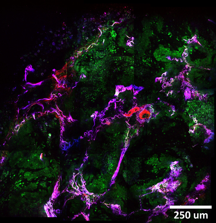
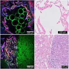
During lung cancer operations a rapid and reliable assessment of tumor tissue can reduce operation time and potentially improve patient outcomes. We show that third harmonic generation (THG), second harmonic generation (SHG) and two-photon excited autofluorescence (2PEF) microscopy reveals relevant, histopathological information within seconds in fresh unprocessed human lung samples. We used a compact, portable microscope and recorded images within 1 to 3 seconds using a power of 5 mW. The generated THG/SHG/2PEF images of tumorous and nontumorous tissues are compared with the corresponding standard histology images, to identify alveolar structures and histopathological hallmarks. Cellular structures (tumor cells, macrophages and lymphocytes) (THG), collagen (SHG) and elastin (2PEF) are differentiated and allowed for rapid identification of carcinoid with solid growth pattern, minimally enlarged monomorphic cell nuclei with salt-and-pepper chromatin pattern, and adenocarcinoma with lipidic and micropapillary growth patterns. THG/SHG/2PEF imaging is thus a promising tool for clinical intraoperative assessment of lung tumor tissue.


 求助内容:
求助内容: 应助结果提醒方式:


