{"title":"ExoHCR:分析肿瘤外泌体上 PD-L1 水平的灵敏检测方法,用于免疫治疗预后。","authors":"Lujun Hu, Wenjie Chen, Shurong Zhou, Guizhi Zhu","doi":"10.1007/s41048-020-00122-x","DOIUrl":null,"url":null,"abstract":"<p><p>Cancer immunotherapy has made recent breakthrough, including immune checkpoint blockade (ICB) that inhibits immunosuppressive checkpoints such as programmed cell death protein 1 (PD-1) and programmed death-ligand 1 (PD-L1). However, most cancer patients do not durably respond to ICB. To predict ICB responses for patient stratification, conventional immunostaining has been used to analyze the PD-L1 expression level on biopsied tumor tissues but has limitations of invasiveness and tumor heterogeneity. Recently, PD-L1 levels on tumor cell exosomes showed the potential to predict ICB response. Here, we developed a non-invasive, sensitive, and fast assay, termed as exosome-hybridization chain reaction (ExoHCR), to analyze tumor cell exosomal PD-L1 levels. First, using αCD63-conjugated magnetic beads, we isolated exosomes from B16F10 melanoma and CT26 colorectal cancer cells that were immunostimulated to generate PD-L1-positive exosomes. Exosomes were then incubated with a conjugate of PD-L1 antibody with an HCR trigger DNA (T), in which one αPD-L1-T conjugate carried multiple copies of T. Next, a pair of metastable fluorophore-labeled hairpin DNA (H1 and H2) were added, allowing T on αPD-L1-T to initiate HCR <i>in situ</i> on bead-conjugated exosome surfaces. By flow cytometric analysis of the resulting beads, relative to αPD-L1-fluorophore conjugates, ExoHCR amplified the fluorescence signal intensities for exosome detection by 3-7 times in B16F10 cells and CT26 cells. Moreover, we validated the biostability of ExoHCR in culture medium supplemented with 50% FBS. These results suggest the potential of ExoHCR for non-invasive, sensitive, and fast PD-L1 exosomal profiling in patient stratification of cancer immunotherapy.</p>","PeriodicalId":59621,"journal":{"name":"生物物理学报:英文版","volume":"6 6","pages":"290-298"},"PeriodicalIF":0.0000,"publicationDate":"2020-12-01","publicationTypes":"Journal Article","fieldsOfStudy":null,"isOpenAccess":false,"openAccessPdf":"https://www.ncbi.nlm.nih.gov/pmc/articles/PMC8320673/pdf/","citationCount":"0","resultStr":"{\"title\":\"ExoHCR: a sensitive assay to profile PD-L1 level on tumor exosomes for immunotherapeutic prognosis.\",\"authors\":\"Lujun Hu, Wenjie Chen, Shurong Zhou, Guizhi Zhu\",\"doi\":\"10.1007/s41048-020-00122-x\",\"DOIUrl\":null,\"url\":null,\"abstract\":\"<p><p>Cancer immunotherapy has made recent breakthrough, including immune checkpoint blockade (ICB) that inhibits immunosuppressive checkpoints such as programmed cell death protein 1 (PD-1) and programmed death-ligand 1 (PD-L1). However, most cancer patients do not durably respond to ICB. To predict ICB responses for patient stratification, conventional immunostaining has been used to analyze the PD-L1 expression level on biopsied tumor tissues but has limitations of invasiveness and tumor heterogeneity. Recently, PD-L1 levels on tumor cell exosomes showed the potential to predict ICB response. Here, we developed a non-invasive, sensitive, and fast assay, termed as exosome-hybridization chain reaction (ExoHCR), to analyze tumor cell exosomal PD-L1 levels. First, using αCD63-conjugated magnetic beads, we isolated exosomes from B16F10 melanoma and CT26 colorectal cancer cells that were immunostimulated to generate PD-L1-positive exosomes. Exosomes were then incubated with a conjugate of PD-L1 antibody with an HCR trigger DNA (T), in which one αPD-L1-T conjugate carried multiple copies of T. Next, a pair of metastable fluorophore-labeled hairpin DNA (H1 and H2) were added, allowing T on αPD-L1-T to initiate HCR <i>in situ</i> on bead-conjugated exosome surfaces. By flow cytometric analysis of the resulting beads, relative to αPD-L1-fluorophore conjugates, ExoHCR amplified the fluorescence signal intensities for exosome detection by 3-7 times in B16F10 cells and CT26 cells. Moreover, we validated the biostability of ExoHCR in culture medium supplemented with 50% FBS. These results suggest the potential of ExoHCR for non-invasive, sensitive, and fast PD-L1 exosomal profiling in patient stratification of cancer immunotherapy.</p>\",\"PeriodicalId\":59621,\"journal\":{\"name\":\"生物物理学报:英文版\",\"volume\":\"6 6\",\"pages\":\"290-298\"},\"PeriodicalIF\":0.0000,\"publicationDate\":\"2020-12-01\",\"publicationTypes\":\"Journal Article\",\"fieldsOfStudy\":null,\"isOpenAccess\":false,\"openAccessPdf\":\"https://www.ncbi.nlm.nih.gov/pmc/articles/PMC8320673/pdf/\",\"citationCount\":\"0\",\"resultStr\":null,\"platform\":\"Semanticscholar\",\"paperid\":null,\"PeriodicalName\":\"生物物理学报:英文版\",\"FirstCategoryId\":\"1089\",\"ListUrlMain\":\"https://doi.org/10.1007/s41048-020-00122-x\",\"RegionNum\":0,\"RegionCategory\":null,\"ArticlePicture\":[],\"TitleCN\":null,\"AbstractTextCN\":null,\"PMCID\":null,\"EPubDate\":\"2020/11/23 0:00:00\",\"PubModel\":\"Epub\",\"JCR\":\"\",\"JCRName\":\"\",\"Score\":null,\"Total\":0}","platform":"Semanticscholar","paperid":null,"PeriodicalName":"生物物理学报:英文版","FirstCategoryId":"1089","ListUrlMain":"https://doi.org/10.1007/s41048-020-00122-x","RegionNum":0,"RegionCategory":null,"ArticlePicture":[],"TitleCN":null,"AbstractTextCN":null,"PMCID":null,"EPubDate":"2020/11/23 0:00:00","PubModel":"Epub","JCR":"","JCRName":"","Score":null,"Total":0}
ExoHCR: a sensitive assay to profile PD-L1 level on tumor exosomes for immunotherapeutic prognosis.
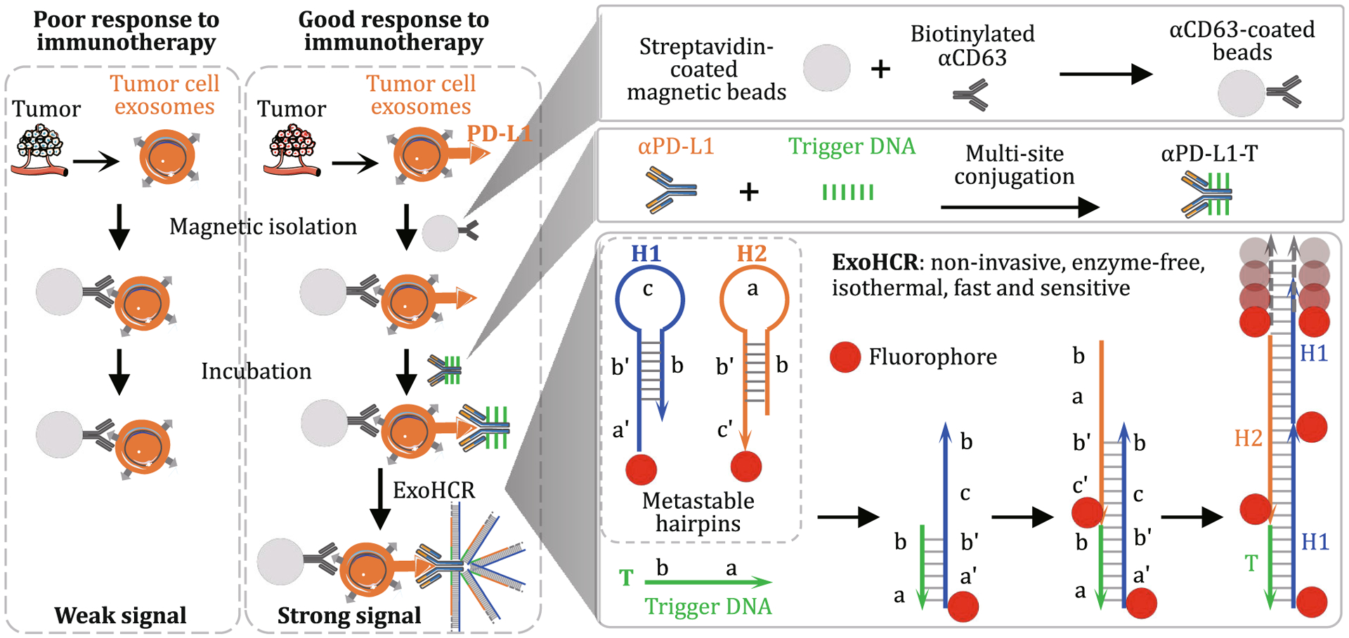
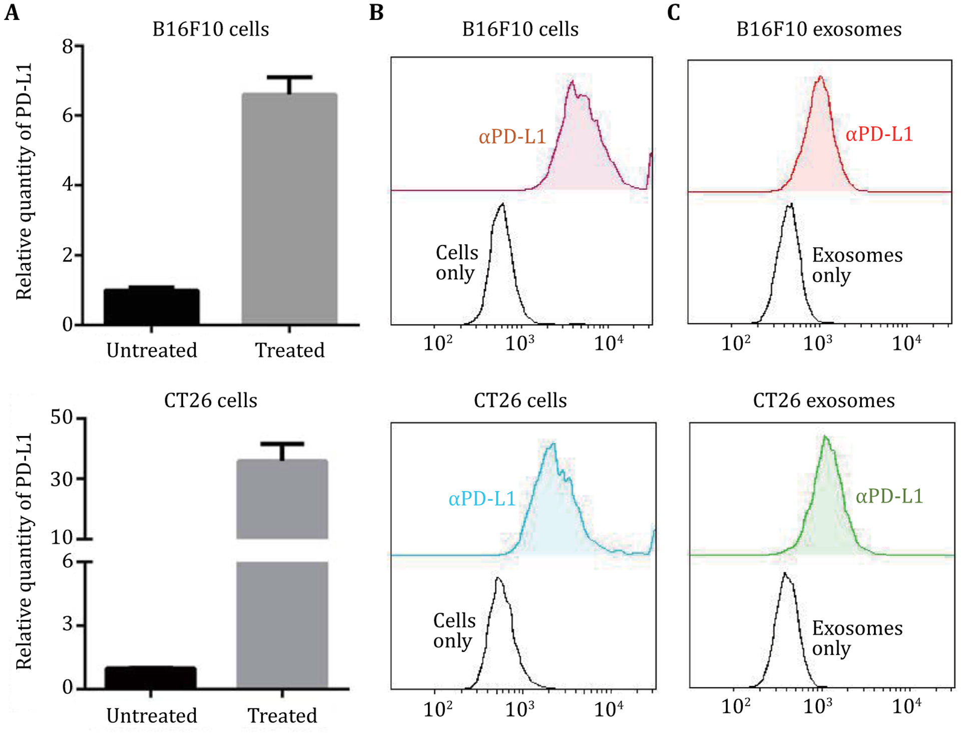
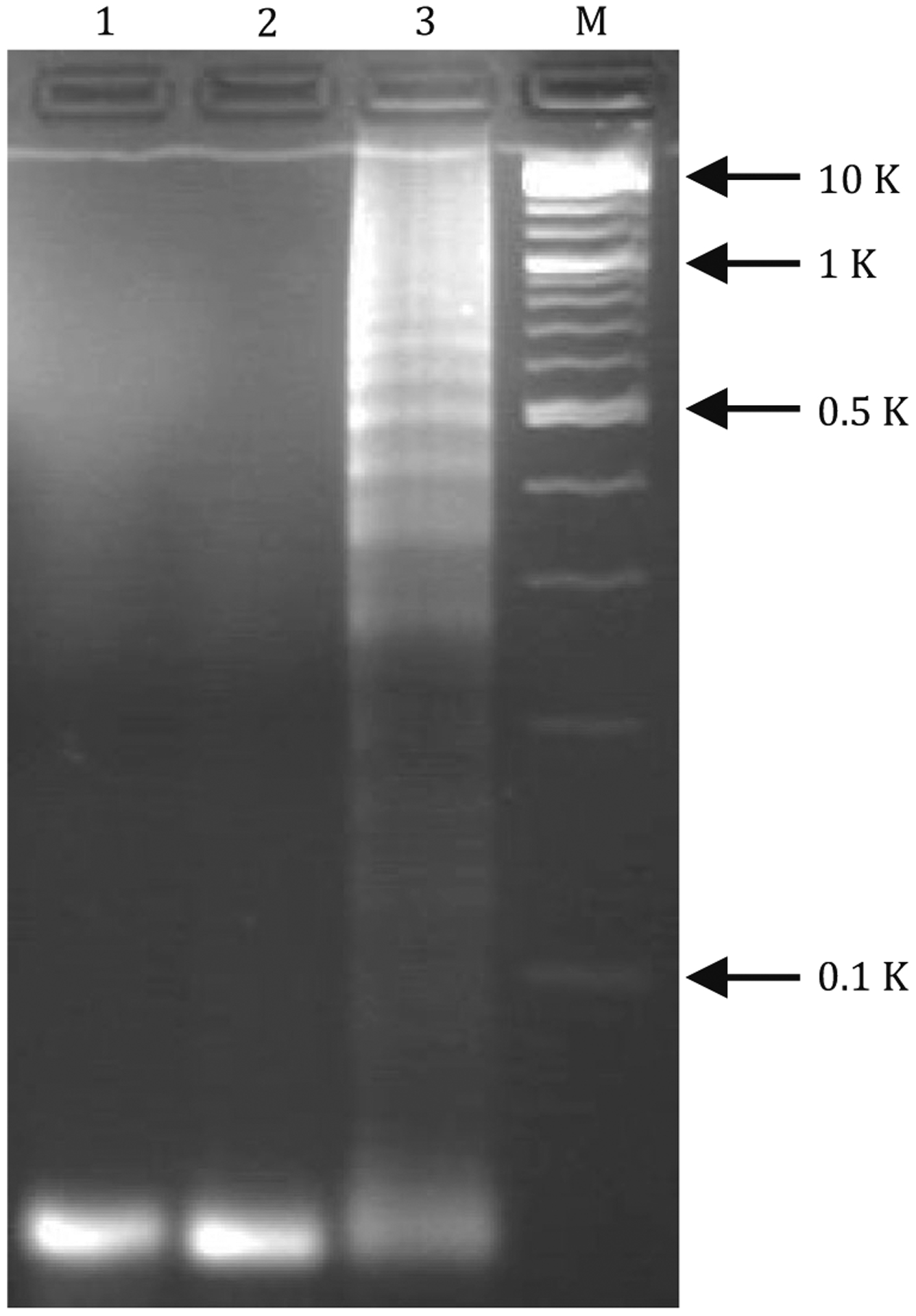
Cancer immunotherapy has made recent breakthrough, including immune checkpoint blockade (ICB) that inhibits immunosuppressive checkpoints such as programmed cell death protein 1 (PD-1) and programmed death-ligand 1 (PD-L1). However, most cancer patients do not durably respond to ICB. To predict ICB responses for patient stratification, conventional immunostaining has been used to analyze the PD-L1 expression level on biopsied tumor tissues but has limitations of invasiveness and tumor heterogeneity. Recently, PD-L1 levels on tumor cell exosomes showed the potential to predict ICB response. Here, we developed a non-invasive, sensitive, and fast assay, termed as exosome-hybridization chain reaction (ExoHCR), to analyze tumor cell exosomal PD-L1 levels. First, using αCD63-conjugated magnetic beads, we isolated exosomes from B16F10 melanoma and CT26 colorectal cancer cells that were immunostimulated to generate PD-L1-positive exosomes. Exosomes were then incubated with a conjugate of PD-L1 antibody with an HCR trigger DNA (T), in which one αPD-L1-T conjugate carried multiple copies of T. Next, a pair of metastable fluorophore-labeled hairpin DNA (H1 and H2) were added, allowing T on αPD-L1-T to initiate HCR in situ on bead-conjugated exosome surfaces. By flow cytometric analysis of the resulting beads, relative to αPD-L1-fluorophore conjugates, ExoHCR amplified the fluorescence signal intensities for exosome detection by 3-7 times in B16F10 cells and CT26 cells. Moreover, we validated the biostability of ExoHCR in culture medium supplemented with 50% FBS. These results suggest the potential of ExoHCR for non-invasive, sensitive, and fast PD-L1 exosomal profiling in patient stratification of cancer immunotherapy.
 求助内容:
求助内容: 应助结果提醒方式:


