{"title":"脑容量测量与0.3和3-T磁共振成像的比较。","authors":"Syo Murata, Akifumi Hagiwara, Hideyoshi Kaga, Yuki Someya, Kiyotaka Nemoto, Masami Goto, Koji Kamagata, Ryusuke Irie, Masaaki Hori, Christina Andica, Akihiko Wada, Kanako Kunishima Kumamaru, Keigo Shimoji, Yujiro Otsuka, Haruyoshi Hoshito, Yoshifumi Tamura, Ryuzo Kawamori, Hirotaka Watada, Shigeki Aoki","doi":"10.2463/mrms.tn.2020-0034","DOIUrl":null,"url":null,"abstract":"<p><p>The volumes of intracranial tissues of 40 healthy volunteers acquired from 0.3- and 3-T scanners were compared using intraclass correlation coefficients, correlation analyses, and Bland-Altman analyses. We found high intraclass correlation coefficients, high Pearson's correlation coefficients, and low percentage biases in all tissues and most of the brain regions, although small differences were observed in some areas. These findings may support the validity of brain volumetry with low-field magnetic resonance imaging.</p>","PeriodicalId":2,"journal":{"name":"ACS Applied Bio Materials","volume":null,"pages":null},"PeriodicalIF":4.6000,"publicationDate":"2022-07-01","publicationTypes":"Journal Article","fieldsOfStudy":null,"isOpenAccess":false,"openAccessPdf":"https://ftp.ncbi.nlm.nih.gov/pub/pmc/oa_pdf/7b/ba/mrms-21-517.PMC9316137.pdf","citationCount":"2","resultStr":"{\"title\":\"Comparison of Brain Volume Measurements Made with 0.3- and 3-T MR Imaging.\",\"authors\":\"Syo Murata, Akifumi Hagiwara, Hideyoshi Kaga, Yuki Someya, Kiyotaka Nemoto, Masami Goto, Koji Kamagata, Ryusuke Irie, Masaaki Hori, Christina Andica, Akihiko Wada, Kanako Kunishima Kumamaru, Keigo Shimoji, Yujiro Otsuka, Haruyoshi Hoshito, Yoshifumi Tamura, Ryuzo Kawamori, Hirotaka Watada, Shigeki Aoki\",\"doi\":\"10.2463/mrms.tn.2020-0034\",\"DOIUrl\":null,\"url\":null,\"abstract\":\"<p><p>The volumes of intracranial tissues of 40 healthy volunteers acquired from 0.3- and 3-T scanners were compared using intraclass correlation coefficients, correlation analyses, and Bland-Altman analyses. We found high intraclass correlation coefficients, high Pearson's correlation coefficients, and low percentage biases in all tissues and most of the brain regions, although small differences were observed in some areas. These findings may support the validity of brain volumetry with low-field magnetic resonance imaging.</p>\",\"PeriodicalId\":2,\"journal\":{\"name\":\"ACS Applied Bio Materials\",\"volume\":null,\"pages\":null},\"PeriodicalIF\":4.6000,\"publicationDate\":\"2022-07-01\",\"publicationTypes\":\"Journal Article\",\"fieldsOfStudy\":null,\"isOpenAccess\":false,\"openAccessPdf\":\"https://ftp.ncbi.nlm.nih.gov/pub/pmc/oa_pdf/7b/ba/mrms-21-517.PMC9316137.pdf\",\"citationCount\":\"2\",\"resultStr\":null,\"platform\":\"Semanticscholar\",\"paperid\":null,\"PeriodicalName\":\"ACS Applied Bio Materials\",\"FirstCategoryId\":\"3\",\"ListUrlMain\":\"https://doi.org/10.2463/mrms.tn.2020-0034\",\"RegionNum\":0,\"RegionCategory\":null,\"ArticlePicture\":[],\"TitleCN\":null,\"AbstractTextCN\":null,\"PMCID\":null,\"EPubDate\":\"2021/7/22 0:00:00\",\"PubModel\":\"Epub\",\"JCR\":\"Q2\",\"JCRName\":\"MATERIALS SCIENCE, BIOMATERIALS\",\"Score\":null,\"Total\":0}","platform":"Semanticscholar","paperid":null,"PeriodicalName":"ACS Applied Bio Materials","FirstCategoryId":"3","ListUrlMain":"https://doi.org/10.2463/mrms.tn.2020-0034","RegionNum":0,"RegionCategory":null,"ArticlePicture":[],"TitleCN":null,"AbstractTextCN":null,"PMCID":null,"EPubDate":"2021/7/22 0:00:00","PubModel":"Epub","JCR":"Q2","JCRName":"MATERIALS SCIENCE, BIOMATERIALS","Score":null,"Total":0}
Comparison of Brain Volume Measurements Made with 0.3- and 3-T MR Imaging.
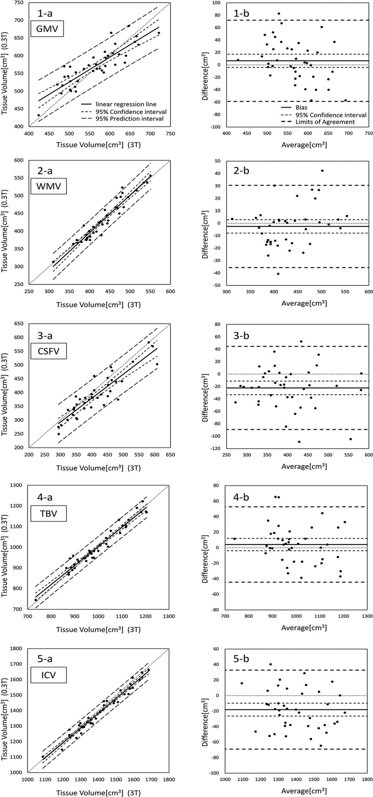
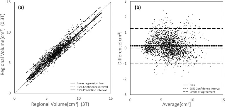
The volumes of intracranial tissues of 40 healthy volunteers acquired from 0.3- and 3-T scanners were compared using intraclass correlation coefficients, correlation analyses, and Bland-Altman analyses. We found high intraclass correlation coefficients, high Pearson's correlation coefficients, and low percentage biases in all tissues and most of the brain regions, although small differences were observed in some areas. These findings may support the validity of brain volumetry with low-field magnetic resonance imaging.