Renata de Lima Bossi, Marcelo Cabral, Monica Oliveira, Sávia Lopes, Rodrigo Hurtado, Marcos Sampaio, Selmo Geber
{"title":"冻干人精子的超微结构分析。","authors":"Renata de Lima Bossi, Marcelo Cabral, Monica Oliveira, Sávia Lopes, Rodrigo Hurtado, Marcos Sampaio, Selmo Geber","doi":"10.5935/1518-0557.20210028","DOIUrl":null,"url":null,"abstract":"<p><strong>Objective: </strong>Lyophilization is potentially more practical and cost-effective alternative for sperm preservation. However, there are no studies that evaluate the ultrastructure of human spermatozoa after lyophilization. Therefore, the aim of our study was to evaluate the ultrasctructure of lyophilized spermatozoa using Transmission Electron Microscopy.</p><p><strong>Methods: </strong>From a total of 21 donated seminal samples, 30 aliquots were originated and divided into two aliquots so that one could have been submitted to cryopreservation/thaw and the other for lyophilization/rehydration. The liquefied aliquots were homogenized at room temperature. Samples assigned for cryopreservation were placed in straws and samples assigned for lyophilization were placed in the appropriate vials. Cryopreservation samples were placed at -30oC for 30 minutes subsequently for 30 minutes at vapour phase and then plunged into liquid nitrogen. Lately, were warmed in water bath at 37oC for 10 minutes followed by 10 minutes centrifugation. The pellet was resuspended and analysed in a Makler chamber. The semen vials assigned for lyophilization were loaded into a pre-fixed freeze-drying chamber. Following lyophilization, vials were removed from the freeze-drying chamber and kept at 4oC until rehydration. TEM was performed after rehydration and thawing. Sperm samples were fixed, rinsed in buffer, post fixed and dehydration was carried out in escalating concentrations of alcohol solution, acetone and then, embedding in Epon resin. Ultrathin sections were stained and examined in a Transmission Electron Microscope.</p><p><strong>Results: </strong>Analysis of sperm after freezing/thawing using Transmission Electron Microscopy showed lesions to the midpiece, with some mitochondria degeneration and random rupture of plasma membrane. In the head, we identified intact plasma membrane, nucleus and acrosome, as in the flagellum all main structures remained intact including the plasma membrane, the longitudinal columns of dense fibers and the semicircular fibers. Analysis by Transmission Electron Microscopy showed that spermatozoa heads had ruptured plasma membranes, absence of acrosomes, nuclei with heterogeneous and decompressed chromatin. Mitochondria were deteriorated in the midpiece. Longitudinal columns of dense fibers were absent in the flagellum. Axonemes, in cross-sections, were disrupted with disorganized structures.</p><p><strong>Conclusions: </strong>To our knowledge, our study demonstrated, for the first time, the structure of the human spermatozoa after lyophilization using Transmission Electron Microscopy. The use of a fixed lyophilization protocol with media containing cryoprotectants might explain the damage to the structures. More studies are necessary to improve the results of sperm lyophilization. In the future, the use of lyophilization of spermatozoa might reduce the costs of fertility preservation, since there will be no need for storage space and transportation is simpler.</p>","PeriodicalId":520656,"journal":{"name":"JBRA assisted reproduction","volume":" ","pages":"473-479"},"PeriodicalIF":1.9000,"publicationDate":"2021-07-21","publicationTypes":"Journal Article","fieldsOfStudy":null,"isOpenAccess":false,"openAccessPdf":"https://www.ncbi.nlm.nih.gov/pmc/articles/PMC8312306/pdf/","citationCount":"5","resultStr":"{\"title\":\"Ultrastructural analysis of Lyophilized Human Spermatozoa.\",\"authors\":\"Renata de Lima Bossi, Marcelo Cabral, Monica Oliveira, Sávia Lopes, Rodrigo Hurtado, Marcos Sampaio, Selmo Geber\",\"doi\":\"10.5935/1518-0557.20210028\",\"DOIUrl\":null,\"url\":null,\"abstract\":\"<p><strong>Objective: </strong>Lyophilization is potentially more practical and cost-effective alternative for sperm preservation. However, there are no studies that evaluate the ultrastructure of human spermatozoa after lyophilization. Therefore, the aim of our study was to evaluate the ultrasctructure of lyophilized spermatozoa using Transmission Electron Microscopy.</p><p><strong>Methods: </strong>From a total of 21 donated seminal samples, 30 aliquots were originated and divided into two aliquots so that one could have been submitted to cryopreservation/thaw and the other for lyophilization/rehydration. The liquefied aliquots were homogenized at room temperature. Samples assigned for cryopreservation were placed in straws and samples assigned for lyophilization were placed in the appropriate vials. Cryopreservation samples were placed at -30oC for 30 minutes subsequently for 30 minutes at vapour phase and then plunged into liquid nitrogen. Lately, were warmed in water bath at 37oC for 10 minutes followed by 10 minutes centrifugation. The pellet was resuspended and analysed in a Makler chamber. The semen vials assigned for lyophilization were loaded into a pre-fixed freeze-drying chamber. Following lyophilization, vials were removed from the freeze-drying chamber and kept at 4oC until rehydration. TEM was performed after rehydration and thawing. Sperm samples were fixed, rinsed in buffer, post fixed and dehydration was carried out in escalating concentrations of alcohol solution, acetone and then, embedding in Epon resin. Ultrathin sections were stained and examined in a Transmission Electron Microscope.</p><p><strong>Results: </strong>Analysis of sperm after freezing/thawing using Transmission Electron Microscopy showed lesions to the midpiece, with some mitochondria degeneration and random rupture of plasma membrane. In the head, we identified intact plasma membrane, nucleus and acrosome, as in the flagellum all main structures remained intact including the plasma membrane, the longitudinal columns of dense fibers and the semicircular fibers. Analysis by Transmission Electron Microscopy showed that spermatozoa heads had ruptured plasma membranes, absence of acrosomes, nuclei with heterogeneous and decompressed chromatin. Mitochondria were deteriorated in the midpiece. Longitudinal columns of dense fibers were absent in the flagellum. Axonemes, in cross-sections, were disrupted with disorganized structures.</p><p><strong>Conclusions: </strong>To our knowledge, our study demonstrated, for the first time, the structure of the human spermatozoa after lyophilization using Transmission Electron Microscopy. The use of a fixed lyophilization protocol with media containing cryoprotectants might explain the damage to the structures. More studies are necessary to improve the results of sperm lyophilization. In the future, the use of lyophilization of spermatozoa might reduce the costs of fertility preservation, since there will be no need for storage space and transportation is simpler.</p>\",\"PeriodicalId\":520656,\"journal\":{\"name\":\"JBRA assisted reproduction\",\"volume\":\" \",\"pages\":\"473-479\"},\"PeriodicalIF\":1.9000,\"publicationDate\":\"2021-07-21\",\"publicationTypes\":\"Journal Article\",\"fieldsOfStudy\":null,\"isOpenAccess\":false,\"openAccessPdf\":\"https://www.ncbi.nlm.nih.gov/pmc/articles/PMC8312306/pdf/\",\"citationCount\":\"5\",\"resultStr\":null,\"platform\":\"Semanticscholar\",\"paperid\":null,\"PeriodicalName\":\"JBRA assisted reproduction\",\"FirstCategoryId\":\"1085\",\"ListUrlMain\":\"https://doi.org/10.5935/1518-0557.20210028\",\"RegionNum\":0,\"RegionCategory\":null,\"ArticlePicture\":[],\"TitleCN\":null,\"AbstractTextCN\":null,\"PMCID\":null,\"EPubDate\":\"\",\"PubModel\":\"\",\"JCR\":\"\",\"JCRName\":\"\",\"Score\":null,\"Total\":0}","platform":"Semanticscholar","paperid":null,"PeriodicalName":"JBRA assisted reproduction","FirstCategoryId":"1085","ListUrlMain":"https://doi.org/10.5935/1518-0557.20210028","RegionNum":0,"RegionCategory":null,"ArticlePicture":[],"TitleCN":null,"AbstractTextCN":null,"PMCID":null,"EPubDate":"","PubModel":"","JCR":"","JCRName":"","Score":null,"Total":0}
Ultrastructural analysis of Lyophilized Human Spermatozoa.
Objective: Lyophilization is potentially more practical and cost-effective alternative for sperm preservation. However, there are no studies that evaluate the ultrastructure of human spermatozoa after lyophilization. Therefore, the aim of our study was to evaluate the ultrasctructure of lyophilized spermatozoa using Transmission Electron Microscopy.
Methods: From a total of 21 donated seminal samples, 30 aliquots were originated and divided into two aliquots so that one could have been submitted to cryopreservation/thaw and the other for lyophilization/rehydration. The liquefied aliquots were homogenized at room temperature. Samples assigned for cryopreservation were placed in straws and samples assigned for lyophilization were placed in the appropriate vials. Cryopreservation samples were placed at -30oC for 30 minutes subsequently for 30 minutes at vapour phase and then plunged into liquid nitrogen. Lately, were warmed in water bath at 37oC for 10 minutes followed by 10 minutes centrifugation. The pellet was resuspended and analysed in a Makler chamber. The semen vials assigned for lyophilization were loaded into a pre-fixed freeze-drying chamber. Following lyophilization, vials were removed from the freeze-drying chamber and kept at 4oC until rehydration. TEM was performed after rehydration and thawing. Sperm samples were fixed, rinsed in buffer, post fixed and dehydration was carried out in escalating concentrations of alcohol solution, acetone and then, embedding in Epon resin. Ultrathin sections were stained and examined in a Transmission Electron Microscope.
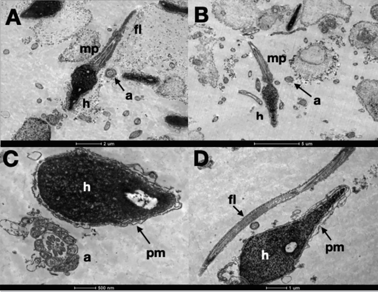
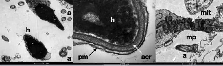
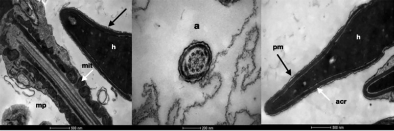
Results: Analysis of sperm after freezing/thawing using Transmission Electron Microscopy showed lesions to the midpiece, with some mitochondria degeneration and random rupture of plasma membrane. In the head, we identified intact plasma membrane, nucleus and acrosome, as in the flagellum all main structures remained intact including the plasma membrane, the longitudinal columns of dense fibers and the semicircular fibers. Analysis by Transmission Electron Microscopy showed that spermatozoa heads had ruptured plasma membranes, absence of acrosomes, nuclei with heterogeneous and decompressed chromatin. Mitochondria were deteriorated in the midpiece. Longitudinal columns of dense fibers were absent in the flagellum. Axonemes, in cross-sections, were disrupted with disorganized structures.
Conclusions: To our knowledge, our study demonstrated, for the first time, the structure of the human spermatozoa after lyophilization using Transmission Electron Microscopy. The use of a fixed lyophilization protocol with media containing cryoprotectants might explain the damage to the structures. More studies are necessary to improve the results of sperm lyophilization. In the future, the use of lyophilization of spermatozoa might reduce the costs of fertility preservation, since there will be no need for storage space and transportation is simpler.

 求助内容:
求助内容: 应助结果提醒方式:


