Rebecca Shi, Daniel A Kramer, Baoyu Chen, Kang Shen
{"title":"两步肌动蛋白聚合机制驱动树突分支。","authors":"Rebecca Shi, Daniel A Kramer, Baoyu Chen, Kang Shen","doi":"10.1186/s13064-021-00154-0","DOIUrl":null,"url":null,"abstract":"<p><strong>Background: </strong>Dendrite morphogenesis plays an essential role in establishing the connectivity and receptive fields of neurons during the development of the nervous system. To generate the diverse morphologies of branched dendrites, neurons use external cues and cell surface receptors to coordinate intracellular cytoskeletal organization; however, the molecular mechanisms of how this signaling forms branched dendrites are not fully understood.</p><p><strong>Methods: </strong>We performed in vivo time-lapse imaging of the PVD neuron in C. elegans in several mutants of actin regulatory proteins, such as the WAVE Regulatory Complex (WRC) and UNC-34 (homolog of Enabled/Vasodilator-stimulated phosphoprotein (Ena/VASP)). We examined the direct interaction between the WRC and UNC-34 and analyzed the localization of UNC-34 in vivo using transgenic worms expressing UNC-34 fused to GFP.</p><p><strong>Results: </strong>We identify a stereotyped sequence of morphological events during dendrite outgrowth in the PVD neuron in C. elegans. Specifically, local increases in width (\"swellings\") give rise to filopodia to facilitate a \"rapid growth and pause\" mode of growth. In unc-34 mutants, filopodia fail to form but swellings are intact. In WRC mutants, dendrite growth is largely absent, resulting from a lack of both swelling and filopodia formation. We also found that UNC-34 can directly bind to the WRC. Disrupting this binding by deleting the UNC-34 EVH1 domain prevented UNC-34 from localizing to swellings and dendrite tips, resulting in a stunted dendritic arbor and reduced filopodia outgrowth.</p><p><strong>Conclusions: </strong>We propose that regulators of branched and linear F-actin cooperate to establish dendritic branches. By combining our work with existing literature, we propose that the dendrite guidance receptor DMA-1 recruits the WRC, which polymerizes branched F-actin to generate \"swellings\" on a mother dendrite. Then, WRC recruits the actin elongation factor UNC-34/Ena/VASP to initiate growth of a new dendritic branch from the swelling, with the help of the actin-binding protein UNC-115/abLIM. Extension of existing dendrites also proceeds via swelling formation at the dendrite tip followed by UNC-34-mediated outgrowth. Following dendrite initiation and extension, the stabilization of branches by guidance receptors further recruits WRC, resulting in an iterative process to build a complex dendritic arbor.</p>","PeriodicalId":49764,"journal":{"name":"Neural Development","volume":" ","pages":"3"},"PeriodicalIF":2.5000,"publicationDate":"2021-07-19","publicationTypes":"Journal Article","fieldsOfStudy":null,"isOpenAccess":false,"openAccessPdf":"https://sci-hub-pdf.com/10.1186/s13064-021-00154-0","citationCount":"9","resultStr":"{\"title\":\"A two-step actin polymerization mechanism drives dendrite branching.\",\"authors\":\"Rebecca Shi, Daniel A Kramer, Baoyu Chen, Kang Shen\",\"doi\":\"10.1186/s13064-021-00154-0\",\"DOIUrl\":null,\"url\":null,\"abstract\":\"<p><strong>Background: </strong>Dendrite morphogenesis plays an essential role in establishing the connectivity and receptive fields of neurons during the development of the nervous system. To generate the diverse morphologies of branched dendrites, neurons use external cues and cell surface receptors to coordinate intracellular cytoskeletal organization; however, the molecular mechanisms of how this signaling forms branched dendrites are not fully understood.</p><p><strong>Methods: </strong>We performed in vivo time-lapse imaging of the PVD neuron in C. elegans in several mutants of actin regulatory proteins, such as the WAVE Regulatory Complex (WRC) and UNC-34 (homolog of Enabled/Vasodilator-stimulated phosphoprotein (Ena/VASP)). We examined the direct interaction between the WRC and UNC-34 and analyzed the localization of UNC-34 in vivo using transgenic worms expressing UNC-34 fused to GFP.</p><p><strong>Results: </strong>We identify a stereotyped sequence of morphological events during dendrite outgrowth in the PVD neuron in C. elegans. Specifically, local increases in width (\\\"swellings\\\") give rise to filopodia to facilitate a \\\"rapid growth and pause\\\" mode of growth. In unc-34 mutants, filopodia fail to form but swellings are intact. In WRC mutants, dendrite growth is largely absent, resulting from a lack of both swelling and filopodia formation. We also found that UNC-34 can directly bind to the WRC. Disrupting this binding by deleting the UNC-34 EVH1 domain prevented UNC-34 from localizing to swellings and dendrite tips, resulting in a stunted dendritic arbor and reduced filopodia outgrowth.</p><p><strong>Conclusions: </strong>We propose that regulators of branched and linear F-actin cooperate to establish dendritic branches. By combining our work with existing literature, we propose that the dendrite guidance receptor DMA-1 recruits the WRC, which polymerizes branched F-actin to generate \\\"swellings\\\" on a mother dendrite. Then, WRC recruits the actin elongation factor UNC-34/Ena/VASP to initiate growth of a new dendritic branch from the swelling, with the help of the actin-binding protein UNC-115/abLIM. Extension of existing dendrites also proceeds via swelling formation at the dendrite tip followed by UNC-34-mediated outgrowth. Following dendrite initiation and extension, the stabilization of branches by guidance receptors further recruits WRC, resulting in an iterative process to build a complex dendritic arbor.</p>\",\"PeriodicalId\":49764,\"journal\":{\"name\":\"Neural Development\",\"volume\":\" \",\"pages\":\"3\"},\"PeriodicalIF\":2.5000,\"publicationDate\":\"2021-07-19\",\"publicationTypes\":\"Journal Article\",\"fieldsOfStudy\":null,\"isOpenAccess\":false,\"openAccessPdf\":\"https://sci-hub-pdf.com/10.1186/s13064-021-00154-0\",\"citationCount\":\"9\",\"resultStr\":null,\"platform\":\"Semanticscholar\",\"paperid\":null,\"PeriodicalName\":\"Neural Development\",\"FirstCategoryId\":\"99\",\"ListUrlMain\":\"https://doi.org/10.1186/s13064-021-00154-0\",\"RegionNum\":3,\"RegionCategory\":\"生物学\",\"ArticlePicture\":[],\"TitleCN\":null,\"AbstractTextCN\":null,\"PMCID\":null,\"EPubDate\":\"\",\"PubModel\":\"\",\"JCR\":\"Q1\",\"JCRName\":\"DEVELOPMENTAL BIOLOGY\",\"Score\":null,\"Total\":0}","platform":"Semanticscholar","paperid":null,"PeriodicalName":"Neural Development","FirstCategoryId":"99","ListUrlMain":"https://doi.org/10.1186/s13064-021-00154-0","RegionNum":3,"RegionCategory":"生物学","ArticlePicture":[],"TitleCN":null,"AbstractTextCN":null,"PMCID":null,"EPubDate":"","PubModel":"","JCR":"Q1","JCRName":"DEVELOPMENTAL BIOLOGY","Score":null,"Total":0}
A two-step actin polymerization mechanism drives dendrite branching.
Background: Dendrite morphogenesis plays an essential role in establishing the connectivity and receptive fields of neurons during the development of the nervous system. To generate the diverse morphologies of branched dendrites, neurons use external cues and cell surface receptors to coordinate intracellular cytoskeletal organization; however, the molecular mechanisms of how this signaling forms branched dendrites are not fully understood.
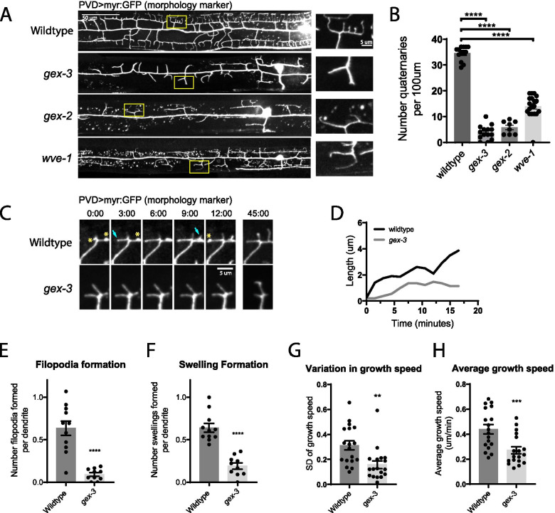
Methods: We performed in vivo time-lapse imaging of the PVD neuron in C. elegans in several mutants of actin regulatory proteins, such as the WAVE Regulatory Complex (WRC) and UNC-34 (homolog of Enabled/Vasodilator-stimulated phosphoprotein (Ena/VASP)). We examined the direct interaction between the WRC and UNC-34 and analyzed the localization of UNC-34 in vivo using transgenic worms expressing UNC-34 fused to GFP.
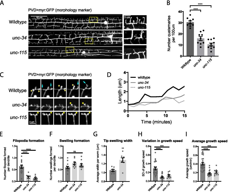
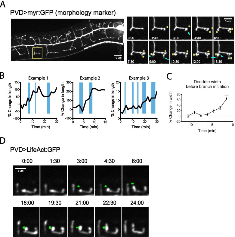
Results: We identify a stereotyped sequence of morphological events during dendrite outgrowth in the PVD neuron in C. elegans. Specifically, local increases in width ("swellings") give rise to filopodia to facilitate a "rapid growth and pause" mode of growth. In unc-34 mutants, filopodia fail to form but swellings are intact. In WRC mutants, dendrite growth is largely absent, resulting from a lack of both swelling and filopodia formation. We also found that UNC-34 can directly bind to the WRC. Disrupting this binding by deleting the UNC-34 EVH1 domain prevented UNC-34 from localizing to swellings and dendrite tips, resulting in a stunted dendritic arbor and reduced filopodia outgrowth.
Conclusions: We propose that regulators of branched and linear F-actin cooperate to establish dendritic branches. By combining our work with existing literature, we propose that the dendrite guidance receptor DMA-1 recruits the WRC, which polymerizes branched F-actin to generate "swellings" on a mother dendrite. Then, WRC recruits the actin elongation factor UNC-34/Ena/VASP to initiate growth of a new dendritic branch from the swelling, with the help of the actin-binding protein UNC-115/abLIM. Extension of existing dendrites also proceeds via swelling formation at the dendrite tip followed by UNC-34-mediated outgrowth. Following dendrite initiation and extension, the stabilization of branches by guidance receptors further recruits WRC, resulting in an iterative process to build a complex dendritic arbor.
期刊介绍:
Neural Development is a peer-reviewed open access, online journal, which features studies that use molecular, cellular, physiological or behavioral methods to provide novel insights into the mechanisms that underlie the formation of the nervous system.
Neural Development aims to discover how the nervous system arises and acquires the abilities to sense the world and control adaptive motor output. The field includes analysis of how progenitor cells form a nervous system during embryogenesis, and how the initially formed neural circuits are shaped by experience during early postnatal life. Some studies use well-established, genetically accessible model systems, but valuable insights are also obtained from less traditional models that provide behavioral or evolutionary insights.
 求助内容:
求助内容: 应助结果提醒方式:


