{"title":"小脑梗死后书写困难1例,功能性近红外光谱引导旨在激活低灌注区的任务。","authors":"Mutsumi Fujii, Kazumi Tanigo, Hirokazu Yamamoto, Keijyu Kikugawa, Masayuki Shirakawa, Miki Ohgushi, Takaaki Chin","doi":"10.1155/2021/6612541","DOIUrl":null,"url":null,"abstract":"<p><strong>Background: </strong>Linguistic impairment following cerebellar lesions is characterized by a marked cerebellocerebral diaschisis with decreased perfusion in the left cerebral hemisphere.</p><p><strong>Case: </strong>We report on a 60-year-old right-handed French chef who presented with linguistic deficits following a right cerebellar infarction. Neurolinguistic examinations in the acute phase showed impaired graphomotor planning, especially for kanji (Japanese morphograms). Despite the absence of any structural damage to the supratentorial brain regions, a quantitative <sup>123</sup>I-IMP SPECT study revealed a relative hypoperfusion, mainly around the left posterior middle temporal gyrus, considered to be a crossed cerebellar-cerebral diaschisis. We performed functional near-infrared spectroscopy (fNIRS) and observed that a picture card task could increase blood perfusion in the affected area. This task was as follows: once he saw a picture card depicting a dish, the patient had to list the ingredients that make up the dish. For example, he had to name vegetables, meat, and spices upon seeing a \"curry\" picture card. We added this task to his daily speech-hearing therapy regimen. In the chronic phase, we confirmed symptom amelioration in linguistic performance-paralleled reduction in the level of hypoperfusion on SPECT study. <i>Discussion</i>. This case is the first report of an fNIRS approach used to evaluate evidence-based prospective speech-hearing tasks by observing blood flow to the hypoperfused area of the cerebral cortex surface.</p>","PeriodicalId":9615,"journal":{"name":"Case Reports in Neurological Medicine","volume":" ","pages":"6612541"},"PeriodicalIF":0.9000,"publicationDate":"2021-06-23","publicationTypes":"Journal Article","fieldsOfStudy":null,"isOpenAccess":false,"openAccessPdf":"https://www.ncbi.nlm.nih.gov/pmc/articles/PMC8249134/pdf/","citationCount":"2","resultStr":"{\"title\":\"A Case of Dysgraphia after Cerebellar Infarction Where Functional NIRS Guided the Task Aimed at Activating the Hypoperfused Region.\",\"authors\":\"Mutsumi Fujii, Kazumi Tanigo, Hirokazu Yamamoto, Keijyu Kikugawa, Masayuki Shirakawa, Miki Ohgushi, Takaaki Chin\",\"doi\":\"10.1155/2021/6612541\",\"DOIUrl\":null,\"url\":null,\"abstract\":\"<p><strong>Background: </strong>Linguistic impairment following cerebellar lesions is characterized by a marked cerebellocerebral diaschisis with decreased perfusion in the left cerebral hemisphere.</p><p><strong>Case: </strong>We report on a 60-year-old right-handed French chef who presented with linguistic deficits following a right cerebellar infarction. Neurolinguistic examinations in the acute phase showed impaired graphomotor planning, especially for kanji (Japanese morphograms). Despite the absence of any structural damage to the supratentorial brain regions, a quantitative <sup>123</sup>I-IMP SPECT study revealed a relative hypoperfusion, mainly around the left posterior middle temporal gyrus, considered to be a crossed cerebellar-cerebral diaschisis. We performed functional near-infrared spectroscopy (fNIRS) and observed that a picture card task could increase blood perfusion in the affected area. This task was as follows: once he saw a picture card depicting a dish, the patient had to list the ingredients that make up the dish. For example, he had to name vegetables, meat, and spices upon seeing a \\\"curry\\\" picture card. We added this task to his daily speech-hearing therapy regimen. In the chronic phase, we confirmed symptom amelioration in linguistic performance-paralleled reduction in the level of hypoperfusion on SPECT study. <i>Discussion</i>. This case is the first report of an fNIRS approach used to evaluate evidence-based prospective speech-hearing tasks by observing blood flow to the hypoperfused area of the cerebral cortex surface.</p>\",\"PeriodicalId\":9615,\"journal\":{\"name\":\"Case Reports in Neurological Medicine\",\"volume\":\" \",\"pages\":\"6612541\"},\"PeriodicalIF\":0.9000,\"publicationDate\":\"2021-06-23\",\"publicationTypes\":\"Journal Article\",\"fieldsOfStudy\":null,\"isOpenAccess\":false,\"openAccessPdf\":\"https://www.ncbi.nlm.nih.gov/pmc/articles/PMC8249134/pdf/\",\"citationCount\":\"2\",\"resultStr\":null,\"platform\":\"Semanticscholar\",\"paperid\":null,\"PeriodicalName\":\"Case Reports in Neurological Medicine\",\"FirstCategoryId\":\"1085\",\"ListUrlMain\":\"https://doi.org/10.1155/2021/6612541\",\"RegionNum\":0,\"RegionCategory\":null,\"ArticlePicture\":[],\"TitleCN\":null,\"AbstractTextCN\":null,\"PMCID\":null,\"EPubDate\":\"2021/1/1 0:00:00\",\"PubModel\":\"eCollection\",\"JCR\":\"Q4\",\"JCRName\":\"CLINICAL NEUROLOGY\",\"Score\":null,\"Total\":0}","platform":"Semanticscholar","paperid":null,"PeriodicalName":"Case Reports in Neurological Medicine","FirstCategoryId":"1085","ListUrlMain":"https://doi.org/10.1155/2021/6612541","RegionNum":0,"RegionCategory":null,"ArticlePicture":[],"TitleCN":null,"AbstractTextCN":null,"PMCID":null,"EPubDate":"2021/1/1 0:00:00","PubModel":"eCollection","JCR":"Q4","JCRName":"CLINICAL NEUROLOGY","Score":null,"Total":0}
A Case of Dysgraphia after Cerebellar Infarction Where Functional NIRS Guided the Task Aimed at Activating the Hypoperfused Region.
Background: Linguistic impairment following cerebellar lesions is characterized by a marked cerebellocerebral diaschisis with decreased perfusion in the left cerebral hemisphere.
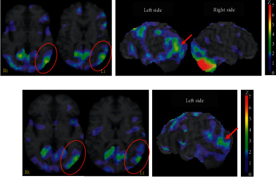
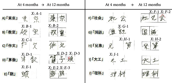
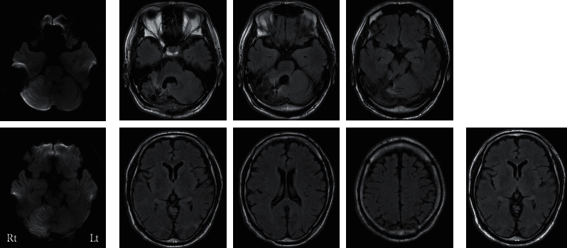
Case: We report on a 60-year-old right-handed French chef who presented with linguistic deficits following a right cerebellar infarction. Neurolinguistic examinations in the acute phase showed impaired graphomotor planning, especially for kanji (Japanese morphograms). Despite the absence of any structural damage to the supratentorial brain regions, a quantitative 123I-IMP SPECT study revealed a relative hypoperfusion, mainly around the left posterior middle temporal gyrus, considered to be a crossed cerebellar-cerebral diaschisis. We performed functional near-infrared spectroscopy (fNIRS) and observed that a picture card task could increase blood perfusion in the affected area. This task was as follows: once he saw a picture card depicting a dish, the patient had to list the ingredients that make up the dish. For example, he had to name vegetables, meat, and spices upon seeing a "curry" picture card. We added this task to his daily speech-hearing therapy regimen. In the chronic phase, we confirmed symptom amelioration in linguistic performance-paralleled reduction in the level of hypoperfusion on SPECT study. Discussion. This case is the first report of an fNIRS approach used to evaluate evidence-based prospective speech-hearing tasks by observing blood flow to the hypoperfused area of the cerebral cortex surface.

 求助内容:
求助内容: 应助结果提醒方式:


