Irim Salik, Nicolas Lamper, Bhupen Mehta, Kar-Mei Chan
{"title":"术中经食管超声心动图监测小儿肝未分化胚胎性肉瘤切除术患者肺栓塞。","authors":"Irim Salik, Nicolas Lamper, Bhupen Mehta, Kar-Mei Chan","doi":"10.1155/2021/5532028","DOIUrl":null,"url":null,"abstract":"<p><p>A minimally invasive monitoring technique, intraoperative transesophageal echocardiography (TEE), has been utilized to provide real-time data on volume status and ventricular function in patients undergoing liver transplantation. In this case, TEE was utilized in an 8-year-old female undergoing undifferentiated embryonal sarcoma of the liver resection to monitor for pulmonary emboli, particularly a saddle embolus. In addition to visualization of cardiac structures, TEE can also be utilized to monitor the liver, lungs, spleen, and kidneys. Monitoring for echocardiographic findings of pulmonary embolism in this high-risk patient was an integral part of effective intraoperative management.</p>","PeriodicalId":36504,"journal":{"name":"Case Reports in Anesthesiology","volume":"2021 ","pages":"5532028"},"PeriodicalIF":0.0000,"publicationDate":"2021-06-19","publicationTypes":"Journal Article","fieldsOfStudy":null,"isOpenAccess":false,"openAccessPdf":"https://www.ncbi.nlm.nih.gov/pmc/articles/PMC8238612/pdf/","citationCount":"0","resultStr":"{\"title\":\"Intraoperative Transesophageal Echocardiography to Monitor for Pulmonary Emboli in a Pediatric Patient Undergoing Undifferentiated Embryonal Sarcoma of the Liver Resection.\",\"authors\":\"Irim Salik, Nicolas Lamper, Bhupen Mehta, Kar-Mei Chan\",\"doi\":\"10.1155/2021/5532028\",\"DOIUrl\":null,\"url\":null,\"abstract\":\"<p><p>A minimally invasive monitoring technique, intraoperative transesophageal echocardiography (TEE), has been utilized to provide real-time data on volume status and ventricular function in patients undergoing liver transplantation. In this case, TEE was utilized in an 8-year-old female undergoing undifferentiated embryonal sarcoma of the liver resection to monitor for pulmonary emboli, particularly a saddle embolus. In addition to visualization of cardiac structures, TEE can also be utilized to monitor the liver, lungs, spleen, and kidneys. Monitoring for echocardiographic findings of pulmonary embolism in this high-risk patient was an integral part of effective intraoperative management.</p>\",\"PeriodicalId\":36504,\"journal\":{\"name\":\"Case Reports in Anesthesiology\",\"volume\":\"2021 \",\"pages\":\"5532028\"},\"PeriodicalIF\":0.0000,\"publicationDate\":\"2021-06-19\",\"publicationTypes\":\"Journal Article\",\"fieldsOfStudy\":null,\"isOpenAccess\":false,\"openAccessPdf\":\"https://www.ncbi.nlm.nih.gov/pmc/articles/PMC8238612/pdf/\",\"citationCount\":\"0\",\"resultStr\":null,\"platform\":\"Semanticscholar\",\"paperid\":null,\"PeriodicalName\":\"Case Reports in Anesthesiology\",\"FirstCategoryId\":\"1085\",\"ListUrlMain\":\"https://doi.org/10.1155/2021/5532028\",\"RegionNum\":0,\"RegionCategory\":null,\"ArticlePicture\":[],\"TitleCN\":null,\"AbstractTextCN\":null,\"PMCID\":null,\"EPubDate\":\"2021/1/1 0:00:00\",\"PubModel\":\"eCollection\",\"JCR\":\"Q3\",\"JCRName\":\"Medicine\",\"Score\":null,\"Total\":0}","platform":"Semanticscholar","paperid":null,"PeriodicalName":"Case Reports in Anesthesiology","FirstCategoryId":"1085","ListUrlMain":"https://doi.org/10.1155/2021/5532028","RegionNum":0,"RegionCategory":null,"ArticlePicture":[],"TitleCN":null,"AbstractTextCN":null,"PMCID":null,"EPubDate":"2021/1/1 0:00:00","PubModel":"eCollection","JCR":"Q3","JCRName":"Medicine","Score":null,"Total":0}
Intraoperative Transesophageal Echocardiography to Monitor for Pulmonary Emboli in a Pediatric Patient Undergoing Undifferentiated Embryonal Sarcoma of the Liver Resection.
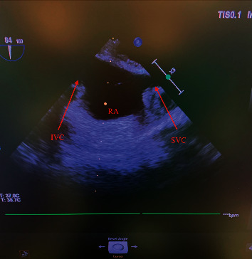
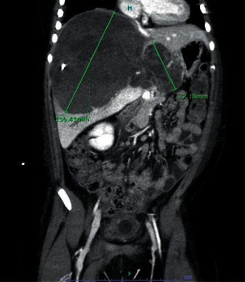
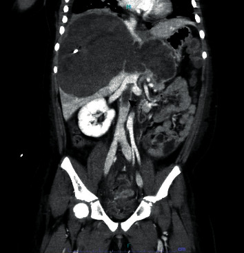
A minimally invasive monitoring technique, intraoperative transesophageal echocardiography (TEE), has been utilized to provide real-time data on volume status and ventricular function in patients undergoing liver transplantation. In this case, TEE was utilized in an 8-year-old female undergoing undifferentiated embryonal sarcoma of the liver resection to monitor for pulmonary emboli, particularly a saddle embolus. In addition to visualization of cardiac structures, TEE can also be utilized to monitor the liver, lungs, spleen, and kidneys. Monitoring for echocardiographic findings of pulmonary embolism in this high-risk patient was an integral part of effective intraoperative management.

 求助内容:
求助内容: 应助结果提醒方式:


