Eike I Piechowiak, Laura Bär, Levin Häni, Mattia Branca, Johannes Kaesmacher, Pasquale Mordasini, Andreas Raabe, Christian T Ulrich, Jan Gralla, Jürgen Beck, Tomas Dobrocky
{"title":"脊髓造影后计算机断层显示肾盂混浊作为自发性颅内低血压患者脑脊液流失的指标。","authors":"Eike I Piechowiak, Laura Bär, Levin Häni, Mattia Branca, Johannes Kaesmacher, Pasquale Mordasini, Andreas Raabe, Christian T Ulrich, Jan Gralla, Jürgen Beck, Tomas Dobrocky","doi":"10.1007/s00062-021-01042-0","DOIUrl":null,"url":null,"abstract":"<p><strong>Purpose: </strong>To assess early renal pelvis opacification on postmyelography computed tomography (CT) as a marker for cerebrospinal fluid (CSF) loss in patients with spontaneous intracranial hypotension (SIH).</p><p><strong>Methods: </strong>The SIH patients referred to our hospital between January 2012 and May 2018 were retrospectively reviewed and divided into 2 groups based on the presence of spinal longitudinal extrathecal CSF collection (SLEC): (1) SLEC(+) with, and (2) SLEC(-) without proof of SLEC on multimodal imaging. Non-SIH patients (n = 20) undergoing CT myelography served as controls. The renal pelvis density on postmyelography CT was measured in all patients. Mean difference in renal pelvis density between the groups was calculated.</p><p><strong>Results: </strong>In total, 111 SIH patients (mean age 48 ± 13 years; 60% female) were included, 71 (64%) SLEC(+) and 40 (36%) SLEC(-). The adjusted renal pelvis density in the SLEC(+), SLEC(-), and the non-SIH group was 108 Hounsfield unit (HU), 83 HU, and 32 HU, respectively, resulting in a significant difference between SLEC(+) vs. control group 1 (75 HU, p < 0.001), SLEC(-) vs. control group 1 (50 HU, p < 0.001), and a tendency for higher density in SLEC(+) than SLEC(-) (25 HU, p = 0.16).</p><p><strong>Conclusion: </strong>Increased renal pelvis opacification on postmyelography CT was observed in SIH patients, even in the absence of a CSF leak or a CSF venous fistula, when compared to non-SIH patients. Although the provenance of early renal opacification in SLEC (-) SIH patients remains unclear, our results suggest that it may be a surrogate for increased spinal CSF resorption via spinal arachnoid granulations and along spinal nerve sheaths occult to direct imaging.</p>","PeriodicalId":49298,"journal":{"name":"Clinical Neuroradiology","volume":"32 2","pages":"529-536"},"PeriodicalIF":2.4000,"publicationDate":"2022-06-01","publicationTypes":"Journal Article","fieldsOfStudy":null,"isOpenAccess":false,"openAccessPdf":"https://sci-hub-pdf.com/10.1007/s00062-021-01042-0","citationCount":"1","resultStr":"{\"title\":\"Renal Pelvis Opacification on Postmyelography Computed Tomography as an Indicator for Cerebrospinal Fluid Loss in Spontaneous Intracranial Hypotension.\",\"authors\":\"Eike I Piechowiak, Laura Bär, Levin Häni, Mattia Branca, Johannes Kaesmacher, Pasquale Mordasini, Andreas Raabe, Christian T Ulrich, Jan Gralla, Jürgen Beck, Tomas Dobrocky\",\"doi\":\"10.1007/s00062-021-01042-0\",\"DOIUrl\":null,\"url\":null,\"abstract\":\"<p><strong>Purpose: </strong>To assess early renal pelvis opacification on postmyelography computed tomography (CT) as a marker for cerebrospinal fluid (CSF) loss in patients with spontaneous intracranial hypotension (SIH).</p><p><strong>Methods: </strong>The SIH patients referred to our hospital between January 2012 and May 2018 were retrospectively reviewed and divided into 2 groups based on the presence of spinal longitudinal extrathecal CSF collection (SLEC): (1) SLEC(+) with, and (2) SLEC(-) without proof of SLEC on multimodal imaging. Non-SIH patients (n = 20) undergoing CT myelography served as controls. The renal pelvis density on postmyelography CT was measured in all patients. Mean difference in renal pelvis density between the groups was calculated.</p><p><strong>Results: </strong>In total, 111 SIH patients (mean age 48 ± 13 years; 60% female) were included, 71 (64%) SLEC(+) and 40 (36%) SLEC(-). The adjusted renal pelvis density in the SLEC(+), SLEC(-), and the non-SIH group was 108 Hounsfield unit (HU), 83 HU, and 32 HU, respectively, resulting in a significant difference between SLEC(+) vs. control group 1 (75 HU, p < 0.001), SLEC(-) vs. control group 1 (50 HU, p < 0.001), and a tendency for higher density in SLEC(+) than SLEC(-) (25 HU, p = 0.16).</p><p><strong>Conclusion: </strong>Increased renal pelvis opacification on postmyelography CT was observed in SIH patients, even in the absence of a CSF leak or a CSF venous fistula, when compared to non-SIH patients. Although the provenance of early renal opacification in SLEC (-) SIH patients remains unclear, our results suggest that it may be a surrogate for increased spinal CSF resorption via spinal arachnoid granulations and along spinal nerve sheaths occult to direct imaging.</p>\",\"PeriodicalId\":49298,\"journal\":{\"name\":\"Clinical Neuroradiology\",\"volume\":\"32 2\",\"pages\":\"529-536\"},\"PeriodicalIF\":2.4000,\"publicationDate\":\"2022-06-01\",\"publicationTypes\":\"Journal Article\",\"fieldsOfStudy\":null,\"isOpenAccess\":false,\"openAccessPdf\":\"https://sci-hub-pdf.com/10.1007/s00062-021-01042-0\",\"citationCount\":\"1\",\"resultStr\":null,\"platform\":\"Semanticscholar\",\"paperid\":null,\"PeriodicalName\":\"Clinical Neuroradiology\",\"FirstCategoryId\":\"3\",\"ListUrlMain\":\"https://doi.org/10.1007/s00062-021-01042-0\",\"RegionNum\":3,\"RegionCategory\":\"医学\",\"ArticlePicture\":[],\"TitleCN\":null,\"AbstractTextCN\":null,\"PMCID\":null,\"EPubDate\":\"2021/6/25 0:00:00\",\"PubModel\":\"Epub\",\"JCR\":\"Q2\",\"JCRName\":\"CLINICAL NEUROLOGY\",\"Score\":null,\"Total\":0}","platform":"Semanticscholar","paperid":null,"PeriodicalName":"Clinical Neuroradiology","FirstCategoryId":"3","ListUrlMain":"https://doi.org/10.1007/s00062-021-01042-0","RegionNum":3,"RegionCategory":"医学","ArticlePicture":[],"TitleCN":null,"AbstractTextCN":null,"PMCID":null,"EPubDate":"2021/6/25 0:00:00","PubModel":"Epub","JCR":"Q2","JCRName":"CLINICAL NEUROLOGY","Score":null,"Total":0}
引用次数: 1
摘要
目的:评价自发性颅内低血压(SIH)患者早期肾盂造影后计算机断层扫描(CT)显示肾盂早期混浊作为脑脊液(CSF)丢失的标志。方法:回顾性分析2012年1月至2018年5月在我院就诊的SIH患者,根据有无脊髓纵向鞘外CSF采集(SLEC)分为2组:(1)SLEC(+), (2) SLEC(-),多模态成像无SLEC证明。非sih患者(n = 20)行CT脊髓造影作为对照。所有患者均在脊髓造影后CT上测量肾盂密度。计算各组肾盂密度的平均差异。结果:共111例SIH患者(平均48岁 ±13岁;其中SLEC(+) 71例(64%),SLEC(-) 40例(36%)。SLEC(+)组、SLEC(-)组和非SIH组调整后的肾盂密度分别为108 Hounsfield单位(HU)、83 HU和32 HU,与对照组1(75 HU, p )相比,SLEC(+)组与对照组1(75 HU, p )有显著差异。结论:与非SIH患者相比,即使没有脑脊液渗漏或脑脊液静脉瘘,SIH患者在脊髓造影后CT上也观察到肾盂浊度增加。尽管SLEC (-) SIH患者早期肾混浊的来源尚不清楚,但我们的研究结果表明,它可能是通过脊髓蛛网膜颗粒和沿直接成像隐蔽的脊神经鞘吸收增加的代用物。
Renal Pelvis Opacification on Postmyelography Computed Tomography as an Indicator for Cerebrospinal Fluid Loss in Spontaneous Intracranial Hypotension.
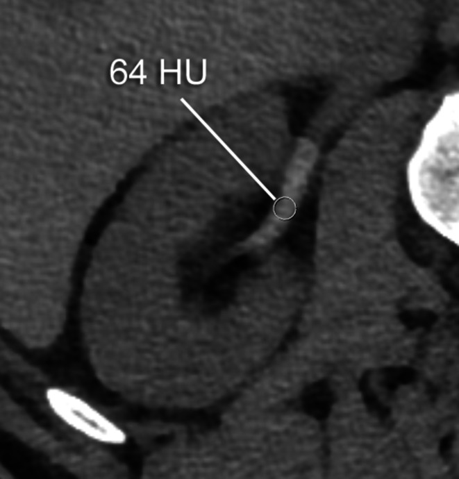
Purpose: To assess early renal pelvis opacification on postmyelography computed tomography (CT) as a marker for cerebrospinal fluid (CSF) loss in patients with spontaneous intracranial hypotension (SIH).
Methods: The SIH patients referred to our hospital between January 2012 and May 2018 were retrospectively reviewed and divided into 2 groups based on the presence of spinal longitudinal extrathecal CSF collection (SLEC): (1) SLEC(+) with, and (2) SLEC(-) without proof of SLEC on multimodal imaging. Non-SIH patients (n = 20) undergoing CT myelography served as controls. The renal pelvis density on postmyelography CT was measured in all patients. Mean difference in renal pelvis density between the groups was calculated.
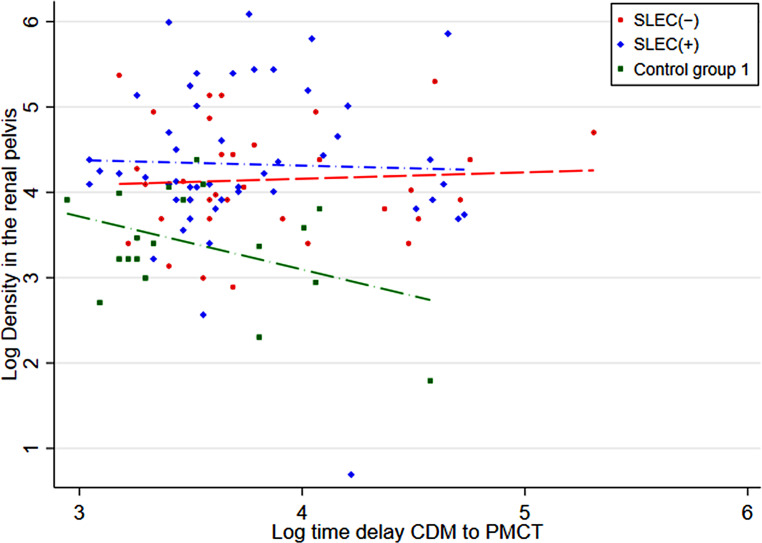
Results: In total, 111 SIH patients (mean age 48 ± 13 years; 60% female) were included, 71 (64%) SLEC(+) and 40 (36%) SLEC(-). The adjusted renal pelvis density in the SLEC(+), SLEC(-), and the non-SIH group was 108 Hounsfield unit (HU), 83 HU, and 32 HU, respectively, resulting in a significant difference between SLEC(+) vs. control group 1 (75 HU, p < 0.001), SLEC(-) vs. control group 1 (50 HU, p < 0.001), and a tendency for higher density in SLEC(+) than SLEC(-) (25 HU, p = 0.16).
Conclusion: Increased renal pelvis opacification on postmyelography CT was observed in SIH patients, even in the absence of a CSF leak or a CSF venous fistula, when compared to non-SIH patients. Although the provenance of early renal opacification in SLEC (-) SIH patients remains unclear, our results suggest that it may be a surrogate for increased spinal CSF resorption via spinal arachnoid granulations and along spinal nerve sheaths occult to direct imaging.
期刊介绍:
Clinical Neuroradiology provides current information, original contributions, and reviews in the field of neuroradiology. An interdisciplinary approach is accomplished by diagnostic and therapeutic contributions related to associated subjects.
The international coverage and relevance of the journal is underlined by its being the official journal of the German, Swiss, and Austrian Societies of Neuroradiology.
 求助内容:
求助内容: 应助结果提醒方式:


