Eyal Klang, Yiftach Barash, Asaf Levartovsky, Noam Barkin Lederer, Adi Lahat
{"title":"基于深度学习的胃溃疡内镜图像良恶性鉴别","authors":"Eyal Klang, Yiftach Barash, Asaf Levartovsky, Noam Barkin Lederer, Adi Lahat","doi":"10.2147/CEG.S292857","DOIUrl":null,"url":null,"abstract":"<p><strong>Background and aim: </strong>Endoscopic differentiation between malignant and benign gastric ulcers (GU) affects further evaluation and prognosis. The aim of our study was to evaluate a deep learning algorithm for discrimination between benign and malignant GU in a database of endoscopic ulcer images.</p><p><strong>Methods: </strong>We retrospectively collected consecutive upper gastrointestinal endoscopy images of GU performed between 2011 and 2019 at the Sheba Medical Center. All ulcers had a corresponding histopathology result of either benign peptic ulcer or gastric adenocarcinoma. A convolutional neural network (CNN) was trained to classify the images into either benign or malignant. Endoscopies from 2011 to 2017 were used for training (2011-2015) and validation (2016-2017). Hyper-parameters, image augmentation and pre-training on Google images obtained images were evaluated on the validation data. Held-out data from 2018 to 2019 was used for testing the final model.</p><p><strong>Results: </strong>Overall, the Sheba dataset included 1978 GU images; 1894 images from benign GU and 84 images of malignant ulcers. The final CNN model showed an AUC 0.91 (95% CI 0.85-0.96) for detecting malignant ulcers. For cut-off probability 0.5, the network showed a sensitivity of 92% and specificity of 75% for malignant ulcers.</p><p><strong>Conclusion: </strong>Our study displays the applicability of a CNN model for automated evaluation of gastric ulcers images for malignant potential. Following further research, the algorithm may improve accuracy of differentiating benign from malignant ulcers during endoscopies and assist in patients' stratification, allowing accelerated patients management and individualized approach towards surveillance endoscopy.</p>","PeriodicalId":10208,"journal":{"name":"Clinical and Experimental Gastroenterology","volume":"14 ","pages":"155-162"},"PeriodicalIF":2.5000,"publicationDate":"2021-05-05","publicationTypes":"Journal Article","fieldsOfStudy":null,"isOpenAccess":false,"openAccessPdf":"https://ftp.ncbi.nlm.nih.gov/pub/pmc/oa_pdf/0f/3a/ceg-14-155.PMC8107004.pdf","citationCount":"10","resultStr":"{\"title\":\"Differentiation Between Malignant and Benign Endoscopic Images of Gastric Ulcers Using Deep Learning.\",\"authors\":\"Eyal Klang, Yiftach Barash, Asaf Levartovsky, Noam Barkin Lederer, Adi Lahat\",\"doi\":\"10.2147/CEG.S292857\",\"DOIUrl\":null,\"url\":null,\"abstract\":\"<p><strong>Background and aim: </strong>Endoscopic differentiation between malignant and benign gastric ulcers (GU) affects further evaluation and prognosis. The aim of our study was to evaluate a deep learning algorithm for discrimination between benign and malignant GU in a database of endoscopic ulcer images.</p><p><strong>Methods: </strong>We retrospectively collected consecutive upper gastrointestinal endoscopy images of GU performed between 2011 and 2019 at the Sheba Medical Center. All ulcers had a corresponding histopathology result of either benign peptic ulcer or gastric adenocarcinoma. A convolutional neural network (CNN) was trained to classify the images into either benign or malignant. Endoscopies from 2011 to 2017 were used for training (2011-2015) and validation (2016-2017). Hyper-parameters, image augmentation and pre-training on Google images obtained images were evaluated on the validation data. Held-out data from 2018 to 2019 was used for testing the final model.</p><p><strong>Results: </strong>Overall, the Sheba dataset included 1978 GU images; 1894 images from benign GU and 84 images of malignant ulcers. The final CNN model showed an AUC 0.91 (95% CI 0.85-0.96) for detecting malignant ulcers. For cut-off probability 0.5, the network showed a sensitivity of 92% and specificity of 75% for malignant ulcers.</p><p><strong>Conclusion: </strong>Our study displays the applicability of a CNN model for automated evaluation of gastric ulcers images for malignant potential. Following further research, the algorithm may improve accuracy of differentiating benign from malignant ulcers during endoscopies and assist in patients' stratification, allowing accelerated patients management and individualized approach towards surveillance endoscopy.</p>\",\"PeriodicalId\":10208,\"journal\":{\"name\":\"Clinical and Experimental Gastroenterology\",\"volume\":\"14 \",\"pages\":\"155-162\"},\"PeriodicalIF\":2.5000,\"publicationDate\":\"2021-05-05\",\"publicationTypes\":\"Journal Article\",\"fieldsOfStudy\":null,\"isOpenAccess\":false,\"openAccessPdf\":\"https://ftp.ncbi.nlm.nih.gov/pub/pmc/oa_pdf/0f/3a/ceg-14-155.PMC8107004.pdf\",\"citationCount\":\"10\",\"resultStr\":null,\"platform\":\"Semanticscholar\",\"paperid\":null,\"PeriodicalName\":\"Clinical and Experimental Gastroenterology\",\"FirstCategoryId\":\"1085\",\"ListUrlMain\":\"https://doi.org/10.2147/CEG.S292857\",\"RegionNum\":0,\"RegionCategory\":null,\"ArticlePicture\":[],\"TitleCN\":null,\"AbstractTextCN\":null,\"PMCID\":null,\"EPubDate\":\"2021/1/1 0:00:00\",\"PubModel\":\"eCollection\",\"JCR\":\"Q2\",\"JCRName\":\"GASTROENTEROLOGY & HEPATOLOGY\",\"Score\":null,\"Total\":0}","platform":"Semanticscholar","paperid":null,"PeriodicalName":"Clinical and Experimental Gastroenterology","FirstCategoryId":"1085","ListUrlMain":"https://doi.org/10.2147/CEG.S292857","RegionNum":0,"RegionCategory":null,"ArticlePicture":[],"TitleCN":null,"AbstractTextCN":null,"PMCID":null,"EPubDate":"2021/1/1 0:00:00","PubModel":"eCollection","JCR":"Q2","JCRName":"GASTROENTEROLOGY & HEPATOLOGY","Score":null,"Total":0}
引用次数: 10
摘要
背景与目的:胃溃疡的内镜鉴别影响进一步的评估和预后。我们研究的目的是评估在内镜溃疡图像数据库中区分良性和恶性GU的深度学习算法。方法:回顾性收集2011年至2019年在示巴医疗中心进行的连续上消化道内镜检查的GU图像。所有溃疡都有相应的组织病理学结果,要么是良性消化性溃疡,要么是胃腺癌。训练卷积神经网络(CNN)将图像分为良性和恶性。2011- 2017年内窥镜用于培训(2011-2015年)和验证(2016-2017年)。在验证数据上对获得的Google图像进行超参数、图像增强和预训练。2018年至2019年的闲置数据用于测试最终模型。结果:Sheba数据集包括1978张GU图像;良性GU 1894张,恶性溃疡84张。最终的CNN模型检测恶性溃疡的AUC为0.91 (95% CI 0.85-0.96)。截止概率为0.5时,该网络对恶性溃疡的敏感性为92%,特异性为75%。结论:我们的研究显示了CNN模型在自动评估胃溃疡图像的恶性潜能方面的适用性。在进一步的研究中,该算法可以提高内镜下溃疡良恶性区分的准确性,并有助于患者分层,加快患者管理和个性化的内镜监测方法。
Differentiation Between Malignant and Benign Endoscopic Images of Gastric Ulcers Using Deep Learning.
Background and aim: Endoscopic differentiation between malignant and benign gastric ulcers (GU) affects further evaluation and prognosis. The aim of our study was to evaluate a deep learning algorithm for discrimination between benign and malignant GU in a database of endoscopic ulcer images.
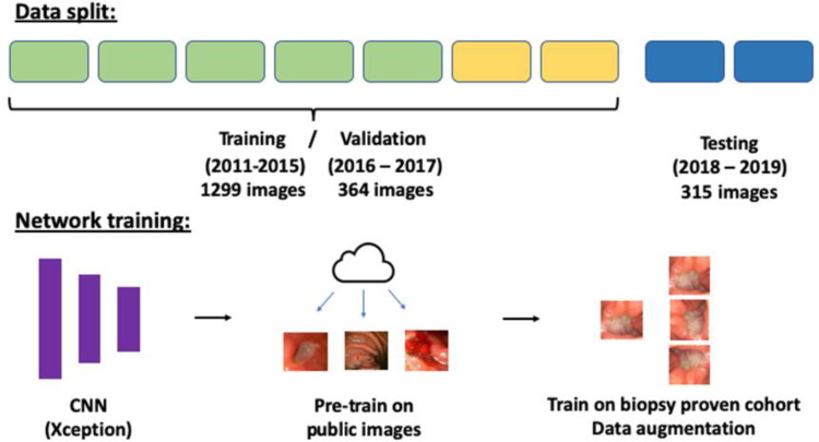
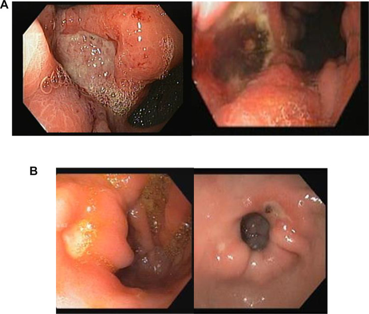
Methods: We retrospectively collected consecutive upper gastrointestinal endoscopy images of GU performed between 2011 and 2019 at the Sheba Medical Center. All ulcers had a corresponding histopathology result of either benign peptic ulcer or gastric adenocarcinoma. A convolutional neural network (CNN) was trained to classify the images into either benign or malignant. Endoscopies from 2011 to 2017 were used for training (2011-2015) and validation (2016-2017). Hyper-parameters, image augmentation and pre-training on Google images obtained images were evaluated on the validation data. Held-out data from 2018 to 2019 was used for testing the final model.
Results: Overall, the Sheba dataset included 1978 GU images; 1894 images from benign GU and 84 images of malignant ulcers. The final CNN model showed an AUC 0.91 (95% CI 0.85-0.96) for detecting malignant ulcers. For cut-off probability 0.5, the network showed a sensitivity of 92% and specificity of 75% for malignant ulcers.
Conclusion: Our study displays the applicability of a CNN model for automated evaluation of gastric ulcers images for malignant potential. Following further research, the algorithm may improve accuracy of differentiating benign from malignant ulcers during endoscopies and assist in patients' stratification, allowing accelerated patients management and individualized approach towards surveillance endoscopy.

 求助内容:
求助内容: 应助结果提醒方式:


