{"title":"明显内淋巴水肿患者的岩上窦截面积减少。","authors":"Shinji Naganawa, Rintaro Ito, Hisashi Kawai, Mariko Kawamura, Toshiaki Taoka, Tadao Yoshida, Michihiko Sone","doi":"10.2463/mrms.mp.2021-0010","DOIUrl":null,"url":null,"abstract":"<p><strong>Purpose: </strong>To evaluate the relationship between the size of the venous structures related to the inner ear and the degree of endolymphatic hydrops (EH).</p><p><strong>Methods: </strong>Thirty-four patients with a suspicion of EH underwent whole brain MR imaging including the inner ear. Images were obtained pre- and post-administration, and at 4 and 24 hours after the intravenous administration of a gadolinium-based contrast agent (IV-GBCA). The cross-sectional areas (CSA) of the internal jugular vein (IJV), superior petrosal sinus (SPS), and inferior petrosal sinus (IPS) were measured on the magnetization prepared rapid acquisition of gradient echo (MPRAGE) images obtained immediately after the IV-GBCA. The grade of EH was determined on the hybrid of reversed image of positive endolymph signal and native image of positive perilymph signal (HYDROPS) images obtained at 4 hours after IV-GBCA as no, mild, and significant EH according to the previously proposed grading system for the cochlea and vestibule, respectively. The ipsilateral CSA was compared between groups with each level of EH grade. P < 0.05 was considered statistically significant.</p><p><strong>Results: </strong>There were no statistically significant differences between EH grades for the CSA of the IJV or that of the IPS in either the cochlea or the vestibule. The CSA of the SPS in the groups with significant EH was significantly smaller than that in the group with no EH, for both the cochlea (P < 0.01) and the vestibule (P < 0.05). In an ROC analysis to predict significant EH, the cut-off CSA value in the SPS was 3.905 mm<sup>2</sup> for the cochlea (AUC: 0.8762, 95% confidence interval [CI]: 0.7952‒0.9572) and 3.805 mm<sup>2</sup> for the vestibule (AUC: 0.7727, 95% CI: 0.6539‒0.8916).</p><p><strong>Conclusion: </strong>In the ears with significant EH in the cochlea or vestibule, the CSA of the ipsilateral SPS was smaller than in the ears without EH.</p>","PeriodicalId":18119,"journal":{"name":"Magnetic Resonance in Medical Sciences","volume":"21 3","pages":"459-467"},"PeriodicalIF":3.2000,"publicationDate":"2022-07-01","publicationTypes":"Journal Article","fieldsOfStudy":null,"isOpenAccess":false,"openAccessPdf":"https://ftp.ncbi.nlm.nih.gov/pub/pmc/oa_pdf/c5/89/mrms-21-459.PMC9316140.pdf","citationCount":"1","resultStr":"{\"title\":\"Cross-sectional Area of the Superior Petrosal Sinus is Reduced in Patients with Significant Endolymphatic Hydrops.\",\"authors\":\"Shinji Naganawa, Rintaro Ito, Hisashi Kawai, Mariko Kawamura, Toshiaki Taoka, Tadao Yoshida, Michihiko Sone\",\"doi\":\"10.2463/mrms.mp.2021-0010\",\"DOIUrl\":null,\"url\":null,\"abstract\":\"<p><strong>Purpose: </strong>To evaluate the relationship between the size of the venous structures related to the inner ear and the degree of endolymphatic hydrops (EH).</p><p><strong>Methods: </strong>Thirty-four patients with a suspicion of EH underwent whole brain MR imaging including the inner ear. Images were obtained pre- and post-administration, and at 4 and 24 hours after the intravenous administration of a gadolinium-based contrast agent (IV-GBCA). The cross-sectional areas (CSA) of the internal jugular vein (IJV), superior petrosal sinus (SPS), and inferior petrosal sinus (IPS) were measured on the magnetization prepared rapid acquisition of gradient echo (MPRAGE) images obtained immediately after the IV-GBCA. The grade of EH was determined on the hybrid of reversed image of positive endolymph signal and native image of positive perilymph signal (HYDROPS) images obtained at 4 hours after IV-GBCA as no, mild, and significant EH according to the previously proposed grading system for the cochlea and vestibule, respectively. The ipsilateral CSA was compared between groups with each level of EH grade. P < 0.05 was considered statistically significant.</p><p><strong>Results: </strong>There were no statistically significant differences between EH grades for the CSA of the IJV or that of the IPS in either the cochlea or the vestibule. The CSA of the SPS in the groups with significant EH was significantly smaller than that in the group with no EH, for both the cochlea (P < 0.01) and the vestibule (P < 0.05). In an ROC analysis to predict significant EH, the cut-off CSA value in the SPS was 3.905 mm<sup>2</sup> for the cochlea (AUC: 0.8762, 95% confidence interval [CI]: 0.7952‒0.9572) and 3.805 mm<sup>2</sup> for the vestibule (AUC: 0.7727, 95% CI: 0.6539‒0.8916).</p><p><strong>Conclusion: </strong>In the ears with significant EH in the cochlea or vestibule, the CSA of the ipsilateral SPS was smaller than in the ears without EH.</p>\",\"PeriodicalId\":18119,\"journal\":{\"name\":\"Magnetic Resonance in Medical Sciences\",\"volume\":\"21 3\",\"pages\":\"459-467\"},\"PeriodicalIF\":3.2000,\"publicationDate\":\"2022-07-01\",\"publicationTypes\":\"Journal Article\",\"fieldsOfStudy\":null,\"isOpenAccess\":false,\"openAccessPdf\":\"https://ftp.ncbi.nlm.nih.gov/pub/pmc/oa_pdf/c5/89/mrms-21-459.PMC9316140.pdf\",\"citationCount\":\"1\",\"resultStr\":null,\"platform\":\"Semanticscholar\",\"paperid\":null,\"PeriodicalName\":\"Magnetic Resonance in Medical Sciences\",\"FirstCategoryId\":\"3\",\"ListUrlMain\":\"https://doi.org/10.2463/mrms.mp.2021-0010\",\"RegionNum\":3,\"RegionCategory\":\"医学\",\"ArticlePicture\":[],\"TitleCN\":null,\"AbstractTextCN\":null,\"PMCID\":null,\"EPubDate\":\"2021/4/24 0:00:00\",\"PubModel\":\"Epub\",\"JCR\":\"Q2\",\"JCRName\":\"RADIOLOGY, NUCLEAR MEDICINE & MEDICAL IMAGING\",\"Score\":null,\"Total\":0}","platform":"Semanticscholar","paperid":null,"PeriodicalName":"Magnetic Resonance in Medical Sciences","FirstCategoryId":"3","ListUrlMain":"https://doi.org/10.2463/mrms.mp.2021-0010","RegionNum":3,"RegionCategory":"医学","ArticlePicture":[],"TitleCN":null,"AbstractTextCN":null,"PMCID":null,"EPubDate":"2021/4/24 0:00:00","PubModel":"Epub","JCR":"Q2","JCRName":"RADIOLOGY, NUCLEAR MEDICINE & MEDICAL IMAGING","Score":null,"Total":0}
Cross-sectional Area of the Superior Petrosal Sinus is Reduced in Patients with Significant Endolymphatic Hydrops.
Purpose: To evaluate the relationship between the size of the venous structures related to the inner ear and the degree of endolymphatic hydrops (EH).
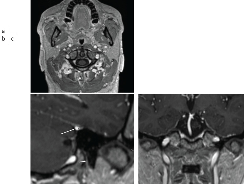
Methods: Thirty-four patients with a suspicion of EH underwent whole brain MR imaging including the inner ear. Images were obtained pre- and post-administration, and at 4 and 24 hours after the intravenous administration of a gadolinium-based contrast agent (IV-GBCA). The cross-sectional areas (CSA) of the internal jugular vein (IJV), superior petrosal sinus (SPS), and inferior petrosal sinus (IPS) were measured on the magnetization prepared rapid acquisition of gradient echo (MPRAGE) images obtained immediately after the IV-GBCA. The grade of EH was determined on the hybrid of reversed image of positive endolymph signal and native image of positive perilymph signal (HYDROPS) images obtained at 4 hours after IV-GBCA as no, mild, and significant EH according to the previously proposed grading system for the cochlea and vestibule, respectively. The ipsilateral CSA was compared between groups with each level of EH grade. P < 0.05 was considered statistically significant.
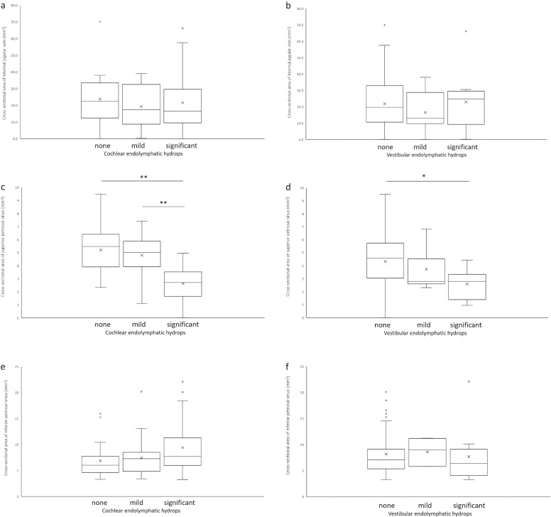
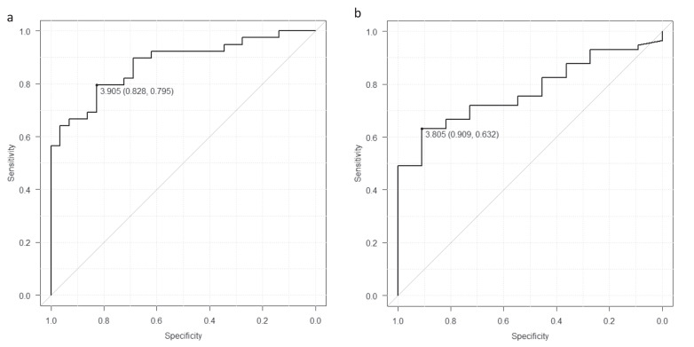
Results: There were no statistically significant differences between EH grades for the CSA of the IJV or that of the IPS in either the cochlea or the vestibule. The CSA of the SPS in the groups with significant EH was significantly smaller than that in the group with no EH, for both the cochlea (P < 0.01) and the vestibule (P < 0.05). In an ROC analysis to predict significant EH, the cut-off CSA value in the SPS was 3.905 mm2 for the cochlea (AUC: 0.8762, 95% confidence interval [CI]: 0.7952‒0.9572) and 3.805 mm2 for the vestibule (AUC: 0.7727, 95% CI: 0.6539‒0.8916).
Conclusion: In the ears with significant EH in the cochlea or vestibule, the CSA of the ipsilateral SPS was smaller than in the ears without EH.
期刊介绍:
Magnetic Resonance in Medical Sciences (MRMS or Magn
Reson Med Sci) is an international journal pursuing the
publication of original articles contributing to the progress
of magnetic resonance in the field of biomedical sciences
including technical developments and clinical applications.
MRMS is an official journal of the Japanese Society for
Magnetic Resonance in Medicine (JSMRM).
 求助内容:
求助内容: 应助结果提醒方式:


