João Paolo Bilibio, Pânila Longhi Lorenzzoni, Brenda Mendes de Oliveira, Flora Leal Nascimento, Arivaldo José Conceição Meireles, Fábio Costa do Nascimento
{"title":"形态学参数、临床因素与整倍体囊胚形成的关系。","authors":"João Paolo Bilibio, Pânila Longhi Lorenzzoni, Brenda Mendes de Oliveira, Flora Leal Nascimento, Arivaldo José Conceição Meireles, Fábio Costa do Nascimento","doi":"10.5935/1518-0557.20210008","DOIUrl":null,"url":null,"abstract":"<p><strong>Objective: </strong>To evaluate the association among embryonic morphological parameters, clinical factors and euploid blastocyst formation.</p><p><strong>Methods: </strong>This prospective cohort study included 422 blastocysts from 135 patients who had undergone preimplantation genetic analysis after intracytoplasmic sperm injection (ICSI).</p><p><strong>Results: </strong>Of 422 blastocysts, 200 (47.4%) were euploid and 222 (52.6%) aneuploid. Women aged older than 38 years were more likely to develop aneuploid embryos (OR: 3.4, CI: 2.2-5.4, p<0.001). Poor ovarian reserve (OR: 3.3, p<0.001), increased male age (39.0 versus 40.7, p=0.019), and decrease in sperm percentage with normal morphology (2.5% vs. 1.9%, p=0.047) were associated with aneuploidy. Type C trophectoderm (TE) and type C inner cell mass were associated with a high risk of embryo aneuploidy, with OR of 4.1 (CI: 2.2-7.7, p<0.001) and 1.7 (CI: 1.01-3.0, p=0.048), respectively. Logistic regression analysis revealed maternal age and type C TE as the main risk factors for aneuploidy. Among combinations of factors, the best marker for the risk of aneuploidy was maternal age older than 38 years, combined with a type-C embryo with trophectoderm, which showed a positive predictive value of 88.6% and a specificity of 97.5%.</p><p><strong>Conclusions: </strong>Trophectoderm and type-C inner cell mass are the main embryo risk factors for aneuploidy, explaining approximately 71% and 60% of the risk, respectively. Among clinical factors, advanced maternal and paternal age (older than 38 and 36 years, respectively), antral follicles (<5), and a low percentage of sperm with normal morphology increased the risk of embryonic aneuploidy.</p>","PeriodicalId":520656,"journal":{"name":"JBRA assisted reproduction","volume":" ","pages":"199-207"},"PeriodicalIF":1.9000,"publicationDate":"2022-04-17","publicationTypes":"Journal Article","fieldsOfStudy":null,"isOpenAccess":false,"openAccessPdf":"https://ftp.ncbi.nlm.nih.gov/pub/pmc/oa_pdf/dd/30/jbra-26-02-0199.PMC9118964.pdf","citationCount":"2","resultStr":"{\"title\":\"Associations among morphological parameters, clinical factors and euploid blastocyst formation.\",\"authors\":\"João Paolo Bilibio, Pânila Longhi Lorenzzoni, Brenda Mendes de Oliveira, Flora Leal Nascimento, Arivaldo José Conceição Meireles, Fábio Costa do Nascimento\",\"doi\":\"10.5935/1518-0557.20210008\",\"DOIUrl\":null,\"url\":null,\"abstract\":\"<p><strong>Objective: </strong>To evaluate the association among embryonic morphological parameters, clinical factors and euploid blastocyst formation.</p><p><strong>Methods: </strong>This prospective cohort study included 422 blastocysts from 135 patients who had undergone preimplantation genetic analysis after intracytoplasmic sperm injection (ICSI).</p><p><strong>Results: </strong>Of 422 blastocysts, 200 (47.4%) were euploid and 222 (52.6%) aneuploid. Women aged older than 38 years were more likely to develop aneuploid embryos (OR: 3.4, CI: 2.2-5.4, p<0.001). Poor ovarian reserve (OR: 3.3, p<0.001), increased male age (39.0 versus 40.7, p=0.019), and decrease in sperm percentage with normal morphology (2.5% vs. 1.9%, p=0.047) were associated with aneuploidy. Type C trophectoderm (TE) and type C inner cell mass were associated with a high risk of embryo aneuploidy, with OR of 4.1 (CI: 2.2-7.7, p<0.001) and 1.7 (CI: 1.01-3.0, p=0.048), respectively. Logistic regression analysis revealed maternal age and type C TE as the main risk factors for aneuploidy. Among combinations of factors, the best marker for the risk of aneuploidy was maternal age older than 38 years, combined with a type-C embryo with trophectoderm, which showed a positive predictive value of 88.6% and a specificity of 97.5%.</p><p><strong>Conclusions: </strong>Trophectoderm and type-C inner cell mass are the main embryo risk factors for aneuploidy, explaining approximately 71% and 60% of the risk, respectively. Among clinical factors, advanced maternal and paternal age (older than 38 and 36 years, respectively), antral follicles (<5), and a low percentage of sperm with normal morphology increased the risk of embryonic aneuploidy.</p>\",\"PeriodicalId\":520656,\"journal\":{\"name\":\"JBRA assisted reproduction\",\"volume\":\" \",\"pages\":\"199-207\"},\"PeriodicalIF\":1.9000,\"publicationDate\":\"2022-04-17\",\"publicationTypes\":\"Journal Article\",\"fieldsOfStudy\":null,\"isOpenAccess\":false,\"openAccessPdf\":\"https://ftp.ncbi.nlm.nih.gov/pub/pmc/oa_pdf/dd/30/jbra-26-02-0199.PMC9118964.pdf\",\"citationCount\":\"2\",\"resultStr\":null,\"platform\":\"Semanticscholar\",\"paperid\":null,\"PeriodicalName\":\"JBRA assisted reproduction\",\"FirstCategoryId\":\"1085\",\"ListUrlMain\":\"https://doi.org/10.5935/1518-0557.20210008\",\"RegionNum\":0,\"RegionCategory\":null,\"ArticlePicture\":[],\"TitleCN\":null,\"AbstractTextCN\":null,\"PMCID\":null,\"EPubDate\":\"\",\"PubModel\":\"\",\"JCR\":\"\",\"JCRName\":\"\",\"Score\":null,\"Total\":0}","platform":"Semanticscholar","paperid":null,"PeriodicalName":"JBRA assisted reproduction","FirstCategoryId":"1085","ListUrlMain":"https://doi.org/10.5935/1518-0557.20210008","RegionNum":0,"RegionCategory":null,"ArticlePicture":[],"TitleCN":null,"AbstractTextCN":null,"PMCID":null,"EPubDate":"","PubModel":"","JCR":"","JCRName":"","Score":null,"Total":0}
Associations among morphological parameters, clinical factors and euploid blastocyst formation.
Objective: To evaluate the association among embryonic morphological parameters, clinical factors and euploid blastocyst formation.
Methods: This prospective cohort study included 422 blastocysts from 135 patients who had undergone preimplantation genetic analysis after intracytoplasmic sperm injection (ICSI).
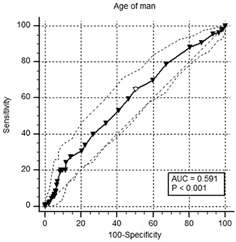
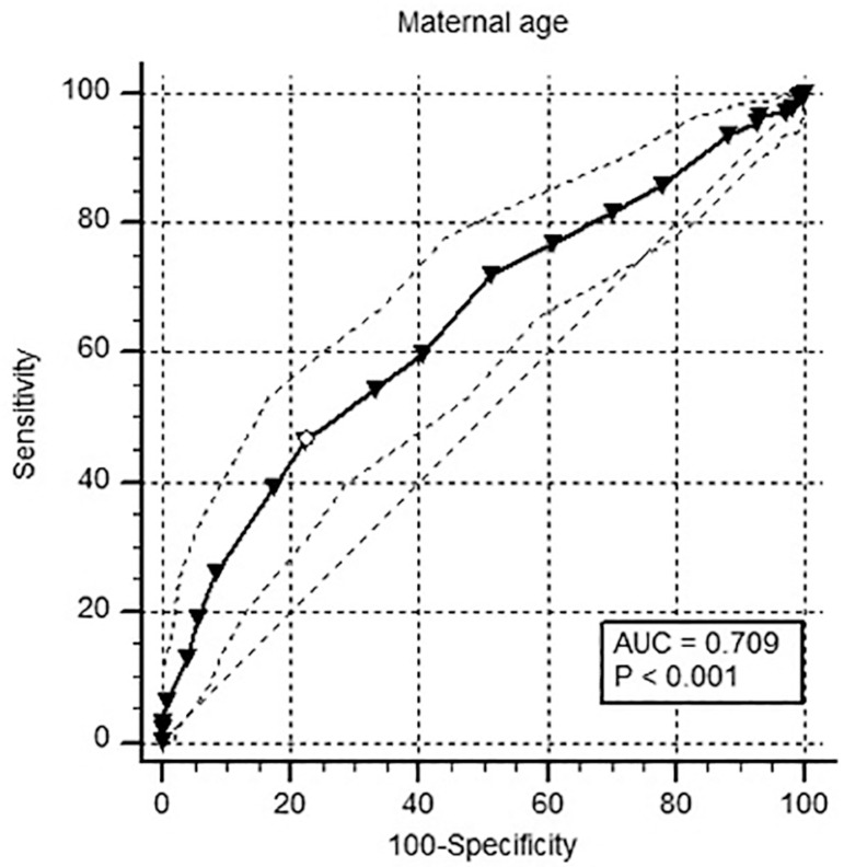
Results: Of 422 blastocysts, 200 (47.4%) were euploid and 222 (52.6%) aneuploid. Women aged older than 38 years were more likely to develop aneuploid embryos (OR: 3.4, CI: 2.2-5.4, p<0.001). Poor ovarian reserve (OR: 3.3, p<0.001), increased male age (39.0 versus 40.7, p=0.019), and decrease in sperm percentage with normal morphology (2.5% vs. 1.9%, p=0.047) were associated with aneuploidy. Type C trophectoderm (TE) and type C inner cell mass were associated with a high risk of embryo aneuploidy, with OR of 4.1 (CI: 2.2-7.7, p<0.001) and 1.7 (CI: 1.01-3.0, p=0.048), respectively. Logistic regression analysis revealed maternal age and type C TE as the main risk factors for aneuploidy. Among combinations of factors, the best marker for the risk of aneuploidy was maternal age older than 38 years, combined with a type-C embryo with trophectoderm, which showed a positive predictive value of 88.6% and a specificity of 97.5%.
Conclusions: Trophectoderm and type-C inner cell mass are the main embryo risk factors for aneuploidy, explaining approximately 71% and 60% of the risk, respectively. Among clinical factors, advanced maternal and paternal age (older than 38 and 36 years, respectively), antral follicles (<5), and a low percentage of sperm with normal morphology increased the risk of embryonic aneuploidy.

 求助内容:
求助内容: 应助结果提醒方式:


